Question
A 45-year-old woman with poorly controlled diabetes mellitus, presented with productive cough for 1 month. She also noticed low grade fever, night sweating and weight loss during this period.
Examination of the chest showed a dull percussion note, decreased air entry and coarse crepitations in the right upper zone.
Laboratory investigations revealed raised ESR, normal white cell count and a CXR was performed.What is the radiological diagnosis?
A. Lung Abscess
B. Empyema
C. Tuberculosis
D. Bronchiectasis
Show Answer
[ads id=”21890″]
|
Correct Answer » C
Explanation
|
|
Ans:C. Tuberculosis
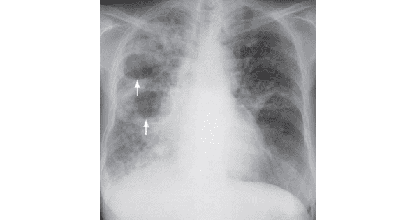
Image shows: Frontal chest radiograph showing air-space opacification in the right lung with cavitating lesions (arrows)
due to caseous necrosis in tuberculosis.
Pulmonary tuberculosis
- The cavitation and air-fluid level within the opacity represents caseous necrosis in tuberculosis.
- The involvement of the apex is a clue towards the diagnosis of TB as post-primary TB has a predilection for the lung apex
Discussion
- Other radiological findings of pulmonary TB not seen in this patient include:
- Enlarged hilum – representing granulomatous inflammation of lymph nodes, usually in primary TB
- Fibrocalcific changes in lung apex usually representing healing of previous TB infection
- Multi-focal air-space opacities representing bronchogenic spread of infection .
- Tiny miliary nodules in both lungs representing miliary TB due to haematogenous spread of infection
Like this:
Like Loading...