Fracture of the facial bones-Nose,Maxilla,Mandible,Zygomatic
Le Forte II facial fracture implies –
| A |
Fracture running through alveolar ridge |
|
| B |
Fracture running through midline of the palate and zygomatico maxillary suture |
|
| C |
Fracture running through zygomatic process of the maxilla, floor of orbit, root of nose on one side only |
|
| D |
Similar to C but on both sides |
Ans. is ‘d’ i.e., Similar to C but on both sides
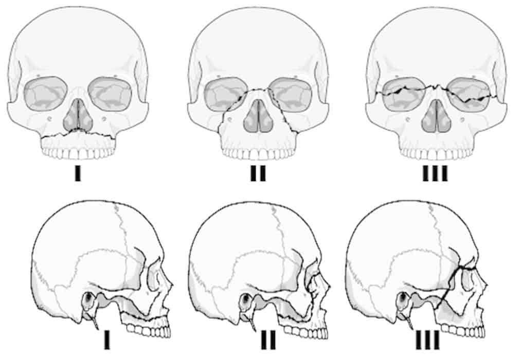
Le Fort Classification of Mid-face #s
- Le Fort I (transverse fracture)
the # line runs above & parallel to the palate and effectively separates the alveolus and palate from the facial skeleton above.
it crosses the lower part of the nasal septum, maxillary antra and the pterygoid plates.
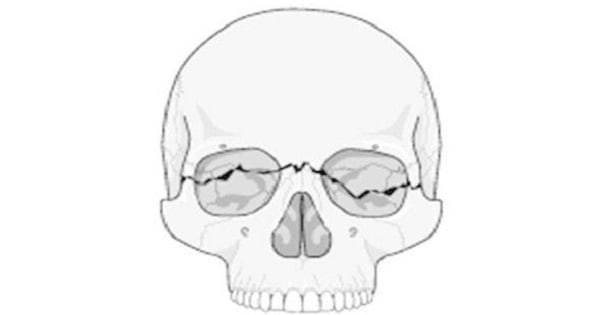
- Le Fort II fracture
it is pyramidal in shape and passes through the root of nose, lacrimal bone, floor of orbit, upper part of maxillary sinus and pterygoid plates. the orbital floor is always involved
- Le Fort III fracture (craniofacial dysjunction) there is complete separation of facial bones from the cranial bones.
the # line runs high through the nasal bridge, septum and ethmoids, and through the bones of orbit to the frontozygomatic suture. The zygomatic
arch fractures and the facial skeleton is separated from the bones above at a high level through the lateral wall of maxillary sinus and the pterygoid plates.
Craniofacial dissociation is seen in:
| A |
Le Fort 1 fracture |
|
| B |
Le Fort 2 fracture |
|
| C |
Le Fort 3 fracture |
|
| D |
Tripod fracture |
Craniofacial dissociation is seen in:
| A |
Le Fort 1 fracture |
|
| B |
Le Fort 2 fracture |
|
| C |
Le Fort 3 fracture |
|
| D |
Tripod fracture |
CSF rhinorrhea is seen in:
| A |
Lefort’s fracture Type I |
|
| B |
Nasal fracture |
|
| C |
Nasoethmoid fracture |
|
| D |
All |
CSF rhinorrhea is seen in:
| A |
Lefort’s fracture Type I |
|
| B |
Nasal fracture |
|
| C |
Nasoethmoid fracture |
|
| D |
All |
CSF Rhinorrhea Occurs in fracture of maxilla in Le Fort type II and type III. (as cribriform plate is injured here) and also in nasal fracture class III
Bone commonly fractured in facial injuries is:
March 2009
| A |
Nasal bones |
|
| B |
Nasoethmoid bone |
|
| C |
Zygomatic bone |
|
| D |
Mandible |
Bone commonly fractured in facial injuries is:
March 2009
| A |
Nasal bones |
|
| B |
Nasoethmoid bone |
|
| C |
Zygomatic bone |
|
| D |
Mandible |
Ans. A: Nasal Bones
Nasal fracture is the most common facial fracture, and the third most common fracture of the skeleton overall. However, because many fractures are subclinical and many others are associated with multiple trauma, a high percentage are not diagnosed or treated at the time of injury.
These often lead to chronic nasal obstruction and account for many of the septoplasty procedures performed for obstruction and septal deviation.
Most facial fractures can be restored to their preoperative state with proper early intervention
Most common site for fracture mandible:
TN 11
| A |
Condyle |
|
| B |
Angle |
|
| C |
Body |
|
| D |
Symphysis |
Most common site for fracture mandible:
TN 11
| A |
Condyle |
|
| B |
Angle |
|
| C |
Body |
|
| D |
Symphysis |
Ans. Condyle
LeFort’s fracture would include all of the following, except:
Manipal 10; WB 09; TN 09; Bihar 10; NIMHANS 14
| A |
Maxilla |
|
| B |
Mandible |
|
| C |
Zygoma |
|
| D |
Nasal bones |
LeFort’s fracture would include all of the following, except:
Manipal 10; WB 09; TN 09; Bihar 10; NIMHANS 14
| A |
Maxilla |
|
| B |
Mandible |
|
| C |
Zygoma |
|
| D |
Nasal bones |
Ans. Mandible
Identify the Type of Le Forte Fracture as shown in the image

| A |
Type II |
|
| B |
Type III |
|
| C |
Type IV |
|
| D |
None of the Above |
Identify the Type of Le Forte Fracture as shown in the image
| A |
Type II |
|
| B |
Type III |
|
| C |
Type IV |
|
| D |
None of the Above |
Fracture mandible occurs most common in ‑
| A |
Body |
|
| B |
Angle |
|
| C |
Condylar process |
|
| D |
Coronoid process |
Fracture mandible occurs most common in ‑
| A |
Body |
|
| B |
Angle |
|
| C |
Condylar process |
|
| D |
Coronoid process |
Ans. is ‘c’ i.e., Condylar process
Condylar process fractures of the mandible are most common account for 35% of all the fractures of mandible. They are followed by angle, body and symphysis in decreasing order of frequency.
Mnemonic CABS: condylar process >angle >body >symphysis decreasing order of frequency of fracture mandible.
Tripod fracture is the name given for
| A |
Zygomatic fracture |
|
| B |
Maxillary fracture |
|
| C |
Mandibular fracture |
|
| D |
Temporal fracture |
Tripod fracture is the name given for
| A |
Zygomatic fracture |
|
| B |
Maxillary fracture |
|
| C |
Mandibular fracture |
|
| D |
Temporal fracture |
Zygoma fracture is also known as tripod fracture.
Clinical features of zygoma fracture
- Considerable swelling over zygomatic arch is common and makes clinical diagnosis more difficult.
- Flattening of malar prominence.
- Step-deformity of infraorbital margin.
- Anaesthesia in the distribution of infraorbital nerve.
- Trismus, due to depression of zygoma on the underlying coronoid process.
- Oblique palpebral fissure, due to the displacement of lateral palpebral ligament.
- Restricted ocular movement, due to entrapment of inferior rectus muscle. It may cause diplopia.
- Periorbital emphysema, due to escape of air from the maxillary sinus on nose-blowing.
- The mucosa of the maxillary sinus may be lacerated and cause epistaxis on that side.
- Fracture of the zygoma may or may not be painful to palpation and running a finger along the zygomatic arch may give a feel of a depressed fracture or a small dimple. The cheek may appear flattened compared to symmetry with the opposite side. This may be obvious immediately following trauma or several days later once swelling has subsided.
Most common site of mandibular fracture is
| A |
Angle of mandible |
|
| B |
Condylar process |
|
| C |
Coronoid process |
|
| D |
Ramus |
Most common site of mandibular fracture is
| A |
Angle of mandible |
|
| B |
Condylar process |
|
| C |
Coronoid process |
|
| D |
Ramus |
Pyramidal fracture of maxilla is
| A |
Le Fort-1 |
|
| B |
Le Fort-2 |
|
| C |
Le Fort-3 |
|
| D |
Craniofacial disruption |
Pyramidal fracture of maxilla is
| A |
Le Fort-1 |
|
| B |
Le Fort-2 |
|
| C |
Le Fort-3 |
|
| D |
Craniofacial disruption |
Fracture of maxilla
It is classified into 3 types : ‑
- Le Fort I (transverse) fracture runs above and parallel to the plate. It crosses lower part of nasal septum, maxillary antra and the pterygoid plates.
- Le Fort II (pyramidal) fracture passes through the root of nose, lacrimal bone, floor of orbit, upper part of maxillary sinus and pterygoid plates. This fracture has some features common with the zygomatic fractures.
- Le Fort III (craniofacial dysjunction). There is complete separation of facial bones from the cranial bones. The fracture line passes through root of nose, ethmofrontal junction, superior orbital fissure, lateral wall of orbit, frontozygomatic and temporozygomatic sutures and the upper part of pterygoid plates.
