fbpx

NEET PG

Palate

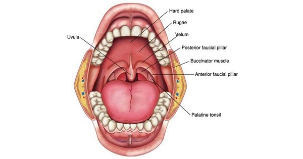
Palate PALATE Palate is the roof of mouth that seperates oral cavity from nasal cavity. It has 2 parts: HARD PALATE( Bony) SOFT PALATE( Muscular) DEVELOPMENT:  Initially, during the 6th week of intrauterine development, there is a common oro-nasal cavity bounded anteriorly by the primary palate and occupied mainly by the developing tongue Formation of […]

Palate Read More »

Palate

PALATE Q. 1 The sensory supply of the palate is through all of the following, EXCEPT?  A Facial nerve  B Hypoglossal nerve  C Glossopharyngeal nerve  D Maxillary division of trigeminal nerve Q. 1 The sensory supply of the palate is through all of the following, EXCEPT?  A Facial nerve  B Hypoglossal nerve  C Glossopharyngeal nerve

Palate Read More »

Foramens of skull

Foramens of skull SKULL: The human skull has numerous holes (foramina) through which cranial nerves, arteries, veins and other structures pass. Nasopalatine nerve Rami nasales posteriores superiores (V2) Bone Cranial fossa Foramina Vessels Nerves Frontal –     Supraorbital foramen Supraorbital artery, supraorbital vein Supraorbital nerve Frontal Anterior cranial fossa     Foramen cecum Emissary veins to superior sagittal sinus from the upper part of

Foramens of skull Read More »

Foramens of skull

FORAMENS OF SKULL Q. 1 Structure passing through Foramen Ovale is?  A Maxillary artery  B Mandibular nerve  C Middle meningeal artery  D Spinal accessory nerve Q. 1 Structure passing through Foramen Ovale is?  A Maxillary artery  B Mandibular nerve  C Middle meningeal artery  D Spinal accessory nerve Ans. B Explanation: Mandibular nerve Q. 2 Structure

Foramens of skull Read More »

Lateral nasal wall

Lateral nasal wall LATERAL NASAL WALL     Has 3 bony projections called as turbinates or conchae. Irregular due to pressence of 3 conchae. Seperates the nose from: Above orbit Below maxillary sinus Front Lacrimal groove & naolacrimal canal Subdivided into: Vestibule small depressed area in ant.  Atrium of middle meatus in middle Posterior contains conchae Bony part: Nasal Frontal process

Lateral nasal wall Read More »

Lateral nasal wall

LATERAL NASAL WALL Q. 1 All open into hiatus semilunaris except  A Posterior ethmoid sinus  B Anterior ethmoid sinus  C Frontal sinus  D Maxillary sinus Q. 1 All open into hiatus semilunaris except  A Posterior ethmoid sinus  B Anterior ethmoid sinus  C Frontal sinus  D Maxillary sinus Ans. A Explanation: Q. 2 Paranasal sinuses communicate

Lateral nasal wall Read More »

Nasal septum/ medial nasal wall

Nasal septum/ medial nasal wall INTRODUCTION: The nasal septum  separates the left and right airways in the nose, dividing the two nostrils. It is depressed by the depressor septi nasi muscle. ANATOMY: Columellar septum It is formed of columella. Containing the medial crura of alar cartilages united together by fibrous tissue. Membranous septum: It lies b/w columella and caudal border of septal cartilage No

Nasal septum/ medial nasal wall Read More »

Acanthamoeba keratitis

Acanthamoeba keratitis ACANTHAMOEBA KERATITIS Acanthamoeba castellani is the most common cause. Does not require a human host to complete the life cycle —free living amoeba In human, acanthamoeba causes: Keratitis Granulamatous encephalitis Fulminant meningoencephalitis Risk factors: Contact lens wearers using home-made saline is the most common cause in developed countries Trauma with contaminated vegetable matter — mcc in

Acanthamoeba keratitis Read More »