

BONES OF HAND- CARPAL
- Short bones, eight in number.
- Arranged in two proximal and distal, each row presenting four bones.
- From lateral to medial side, bones of proximal row are scaphoid, lunate, triquetrum and pisiform.
- bones of distal row are trapezium, trapezoid, capitate and hamate.
OSSIFICATION:
- Ossification of carpal bones appear after birth in a spiral manner as follows:
|
CARPAL BONE |
OSSIFICATION (in yrs) |
|
Capitate |
1 |
|
Hamate |
2 |
|
Triquetrum |
3 |
|
Lunate |
4 |
|
Scaphoid |
5 |
|
Trapezium |
6 |
|
Trapezoid |
7 |
|
Pisiform |
12 |

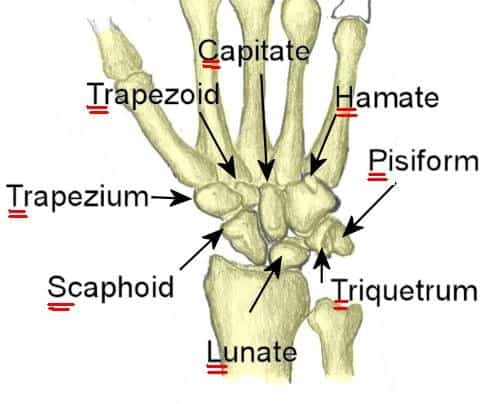
Note: The scaphoid bone is a small carpal bone on the thumb side (radial side) of the wrist. It is the most commonly fractured carpal bone.
- This is probably because it actually crosses two rows of carpal bones, forming a hinge.
Articulations:
|
CARPAL BONE |
ARTICULATING WITH |
|
Scaphoid |
Radius, trapezium, trapezoid, lunate, capitate |
|
Lunate |
Radius, capitate, hamate, scaphoid, Triquetral |
|
Triquetrum |
Hamate, lunate, pisiform |
|
Pisiform |
Triquetral Minimum articulation with only 1 bone |
|
Trapezium |
Scaphoid, trapezoid, 1st & 2nd metacarpal |
|
Trapezoid |
Scaphoid, trapezium, capitate, 2nd metacarpal |
|
Capitate |
Scaphoid, lunate, trapezoid, hamate, 2nd 3rd & 4th metacarpals Maximum articulation with 7 bones |
|
Hamate |
Lunate, capitate, Triquetral, 4th & 5th metacarpals |
BONY PILLARS:
- There are four bony pillars at the four corners of the carpus.
- All attachments are to these four pillars:
- The flexor retinaculum
- A few fibers of the abductor pollicis brevis
- Flexor carpi ulnaris
- Flexor retinaculum and its superficial slip
- Abductor digiti minimi
- Extensor retinaculum
- The crest gives origin to the abductor pollicis brevis, flexor pollicis brevis, and opponens pollicis. These constitute muscles of thenar eminence.
- The edges of the groove give attachment to the two layers of the flexor retinaculum.
- The lateral surface gives attachment to the lateral ligament of the wrist joint.
- The groove lodges the tendon of the flexor carpi radialis.
- The tip of the hook gives attachment to the flexor retinculum.
- The medial side of the hook gives attachment to the flexor digiti minimi and the opponens digiti minimi.
– Kienbock’s disease is osteochondritis of lunate.
– Capitate is the largest carpal bone, first carpal bone to ossify and articulates with maximum number of bones.
– Lunate is the most commonly dislocated carpal bone.
– Scaphoid is the most commonly fractured carpal bone.
- The scaphoid bone is a small carpal bone on the thumb side (radial side) of the wrist. It is the most commonly fractured carpal bone.
- This is probably because it actually crosses two rows of carpal bones, forming a hinge.
Exam Important
- 4th carpal bones (Lunate) are present at 4 yrs of age.
- Carpal bones articulate with radius are Scaphoid and lunate.
- Carpal bone which fracture commonly is Scaphoid.
- The commonly injured carpal bone next to scaphoid is Trapezium.
- Lunate carpal bone fracture causes median nerve involvement.
Click Here to Start Quiz

