
Cellular adaptation

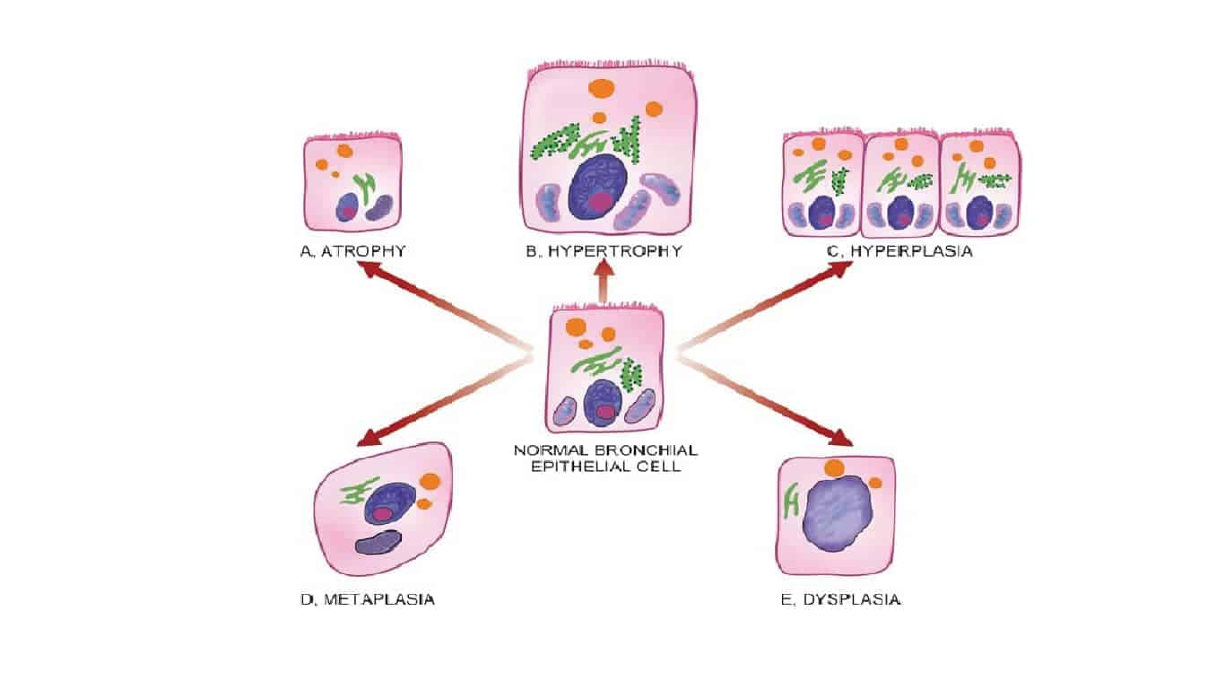
- Adaptations are reversible changes in the cell which can be physiologic (normal stimulation by hormones or mediators) or pathological (stress changes structure & function). It includes-
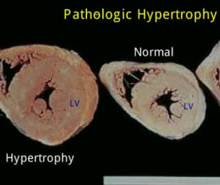
1. Hypertrophy-
- Hypertrophy is an increase in the size of cells resulting in increase in the size of the organ.
- There is no change in the number of cells.
- Hypertrophy can be of two types-
Physiologic
- Enlarged size of uterus in pregnancy, breast tissue in puberty & pregnancy is an example of hypertrophy as well as hyperplasia.


Pathological
- Tissues showing hypertrophy in cardiac muscles (left ventricular hypertrophy) , smooth muscle, skeletal muscle.

2. Hyperplasia-
- Hyperplasia is an increase in the number of cells but there is no change in the size of tissue.
- Hyperplasia takes place in cells, which are capable of synthesizing DNA.
- Hyperplasia can be-
a) Physiologic- 2 types
- Hormonal- Examples are- female breast at puberty, pregnancy and pregnant uterus.
- Compensatory- E. g.- regeneration of liver, epidermis.
b) Pathological- Endometrial hyperplasia, wound healing, BPH.


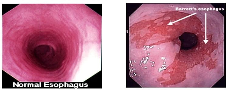
3. Metaplasia-
- Metaplasia is a reversible change in which one adult cell type (epithelial or mesenchymal) is replaced by another adult cell type.
- It is due to re- programming of stem cells.
- Vitamin A deficiency (in respiratory epithelium) or excess leads to metaplasia.
- Metaplasia is of 2 types-
a) Epithelial- It is of two types
i) Squamous- examples are
- Uterine endocervix in prolapsed uterus & old age.
- Gallbladder, salivary gland, pancreas in chronic cholecystitis with cholelithiasis.
- Bronchus in chronic smoker.
- Connective tissue metaplasia
- bone formation in muscle (myositis ossificans)
ii) Columnar – Columnar metaplasia in Barrett’s oesophagus (intestinal metaplasia).

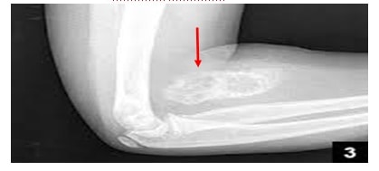
b) Mesenchymal-
- Osseous-
1. In soft tissues in myositis ossificans.

2. In arterial wall in old age.
ii) Cartilaginous- Healing of fractures.
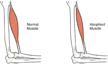
4. Atrophy-
- Both number and size of cells are decreased.
- Mechanism of atrophy consists of combination of decreased protein synthesis & increased protein degradation by Ubiquitin proteasome pathway.
- Lysosomal acid hydrolases
Types-
a) Physiological atrophy- eg. atrophy of notochord or thyroglossal duct during fetal development and uterus after parturition.
b) Pathologial atrophy-
Eg. In denervation atrophy
Atherosclerosis can cause ischaemic atrophy
Nutritional deficiency, eg, marasmus and cancer cachexia (nutritional atrophy)
Senile atrophy is an ageing-associated eg. brain and heart or testes

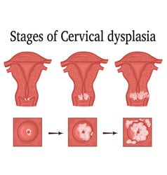
5. Dysplasia-
- Dysplasia is disordered cellular development.
- It affects only epithelial cells.
- Most common examples are uterine cervix and respiratory tract.

Exam Important
- Hypertrophy is an increase in the size of cells resulting in increase in the size of the organ and there is no change in the number of cells.
- Hypertrophy examples- Physiologic –
1. Enlarged size of uterus in pregnancy, breast tissue in puberty & pregnancy is an example of hypertrophy as well as hyperplasia.
- Hyperplasia– Hyperplasia is an increase in the number of cells but there is no change in the size of tissue.
- Hyperplasia examples- Physiologic (Hormonal)- female breast at puberty, pregnancy and pregnant uterus.
- Metaplasia– Vitamin A deficiency (in respiratory epithelium) or excess leads to metaplasia.
- Metaplasia can be two types-
Epithelial-
- Squamous- Uterine endocervix in prolapsed uterus & old age, Bronchus in chronic smoker.
- Columnar- Columnar metaplasia in Barrett’s oesophagus (intestinal metaplasia).
2. Mesenchymal example is myositis ossificans
- Mechanism of atrophy is by Ubiquitin proteasome pathway.
Click Here to Start Quiz

