

Question
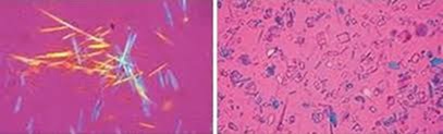
A patient presents with knee joint pain and swelling. Synovial fluid analysis reveals positively birefringent rhomboid-shaped crystals under polarized light microscopy (image shown). What is the most likely diagnosis?

| A. |
Gout |
| B. |
Pseudogout |
| C. |
Septic arthritis |
| D. |
Rheumatoid arthritis |


|
Correct Answer » B Explanation |
 |

– Positively birefringent rhomboid crystals → calcium pyrophosphate dihydrate (CPPD) crystals.
– Seen in pseudogout, also called Calcium Pyrophosphate Deposition Disease (CPPD).
– Crystals appear blue when parallel to the polarizer.
– Most commonly affects the knee & occurs in the elderly.
– Associated with hyperparathyroidism, hemochromatosis, hypomagnesemia, hypothyroidism.
– Presents as acute monoarthritis, mimicking gout or septic arthritis.
Incorrect Options:
A. Gout – Monosodium urate crystals show negative birefringence & needle shape.
C. Septic arthritis – High WBCs, neutrophils dominate, no crystals.
D. Rheumatoid arthritis – Chronic polyarthritis, inflammatory fluid, no crystals.