
Question
Identify the nerve block shown in the given image:
| A. | Inferior alveolar |
| B. |
Anterior ethmoidal |
| C. |
Maxillary |
| D. |
Greater palatine |

|
Correct Answer � D Explanation |
|

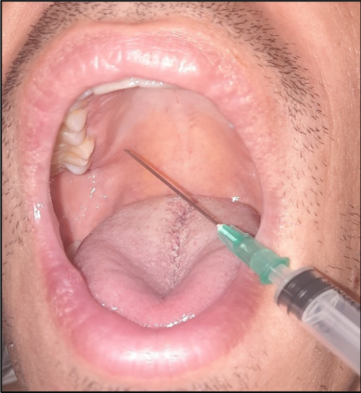
Image Interpretation:
– Needle inserted into posterior hard palate near 2nd/3rd molar.
– Located close to greater palatine foramen.
– Typical site for greater palatine nerve block.
Anatomy:
– Greater palatine nerve: branch of maxillary nerve (V2).
– Exits via greater palatine foramen, medial to 3rd maxillary molar.
– Supplies posterior hard palate & palatal gingiva up to canine.
Clinical Use:
– Provides palatal anesthesia for:
• Posterior palatal soft tissue procedures,
• Posterior teeth extraction,
• Palatal surgeries.
Why Other Options Are Incorrect:
A. Inferior alveolar nerve block:
• Given near mandibular foramen.
• Anesthetizes mandibular teeth & lower lip, not palate.
B. Anterior ethmoidal nerve block:
• Nasal block, unrelated to oral cavity anesthesia.
C. Maxillary nerve block:
• Given via pterygopalatine fossa or infrazygomatic approach.
• Not administered on the palate directly.