Question
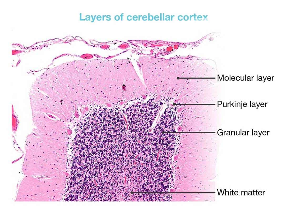
Identify the structures marked as A, B, C, and D in the histological image below the cerebellar cortex.


| A. |
A= Molecular layer, B = Purkinje layer, C = Granular layer, D = White matter |
| B. |
A=White matter, B = Purkinje layer, C = Granular layer, D = Molecular layer |
| C. |
A= White matter, B = Granular layer, C = Purkinje layer, D = Molecular layer |
| D. |
A= Molecular layer, B = Granular layer, C = Purkinje layer, D = White matter |
Show Answer


|
Correct Answer � A Explanation |
 |

The structures marked in the given image are:
A – Molecular layer
B – Purkinje layer
C – Granular layer
D – White matter
The cerebellum is covered by pia mater which is seen as thin connective tissue, with blood vessels lying under it. The cerebellum consists of an outer grey matter (cerebellar cortex) and inner white matter.