

Question

| A. | Spironolactone |
| B. |
Thiazides |
| C. |
Prazosin |
| D. |
Metoprolol |


|
Correct Answer » A Explanation |
 |

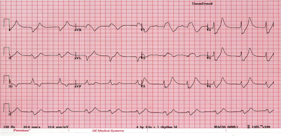
The Above ECG Shows
– Tall Peaked T Waves
– Loss of P Waves
The Above changes can be correlated with hyperkalemia typically seen with use of spironolactone.
Each of the drugs mentioned can have a different impact on cardiovascular physiology and thus on the electrocardiogram (ECG) findings. Here are the potential ECG changes or considerations associated with each:
Spironolactone: Hyperkalemia: Being a potassium-sparing diuretic, spironolactone can lead to elevated potassium levels. ECG changes associated with hyperkalemia include:
– Tall peaked T waves
– Prolonged PR interval and QRS duration
– Loss of P wave
– Sine wave appearance in severe cases
– Ventricular fibrillation or asystole in very severe cases
Thiazides:
Hypokalemia: Thiazides can lead to potassium loss. ECG changes associated with hypokalemia include:
– ST segment depression
– T wave flattening or inversion
– U wave prominence (a wave after the T wave)
Hyponatremia: Sodium levels can also decrease with thiazide use, but specific ECG changes are less distinctive. Severe hyponatremia can increase the risk of other cardiac arrhythmias.
Prazosin: Prazosin is an alpha-1 blocker and is primarily used for hypertension and benign prostatic hyperplasia.
Direct ECG changes are not commonly associated with prazosin. However, it can cause postural (orthostatic) hypotension, especially after the first dose. While postural hypotension itself does not cause specific ECG changes, it’s crucial to be aware of this side effect clinically.
Metoprolol: Metoprolol is a beta-blocker and can cause the following ECG changes:
– Bradycardia: Reduced heart rate is a common side effect.
– Prolonged PR interval: Beta-blockers can delay AV nodal conduction.
– Possible masking of hypoglycemia-induced tachycardia: In diabetics, hypoglycemia usually causes tachycardia as a symptom, but beta-blockers like metoprolol can mask this sign.