Question

| A. |
Pancreatic calcification |
| B. |
Mesenteric calcification |
| C. |
Horseshoe kidney |
| D. |
Jejunal fecolith |


|
Correct Answer » A Explanation |
 |

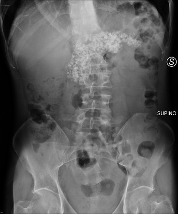
The radiograph shows calcifications located in the midline, consistent with pancreatic calcifications. Pancreatic calcification is often associated with chronic pancreatitis, where long-standing inflammation leads to the deposition of calcium within the pancreatic tissue.
Why the Other Options Are Incorrect:
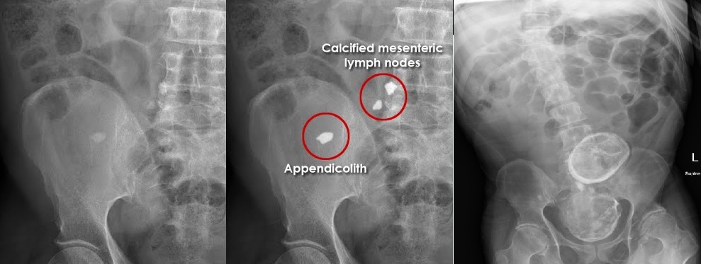
Mesenteric calcification: Refers to calcifications within the mesentery, often due to conditions like mesenteric lymphadenitis or mesenteric ischemia, but typically appears in a different pattern and location.
Horseshoe kidney: A congenital condition where the kidneys are fused at the lower poles, forming a U-shape. It does not present with calcifications in this pattern.
Jejunal fecolith: Refers to a stone-like mass of hardened stool in the jejunum, which would not produce the calcification pattern seen here.