

Question
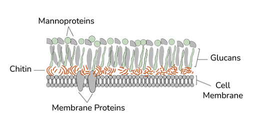
The cell wall depicted below is most likely to belong to which of the following organisms?

| A. |
Rhinosporidium seeberi |
| B. |
Mycobacterium tuberculosis |
| C. |
Mycoplasma pneumoniae |
| D. |
Aspergillus fumigatus |
Show Answer


|
Correct Answer » D Explanation |
 |

- The diagram clearly shows:
- Outer layer: Mannoproteins
- Middle layer: β-glucans
- Inner layer: Chitin
- This layered structure is typical of fungal cell walls
Aspergillus fumigatus
- A fungus (mold)
- Cell wall composition:
- Chitin (structural rigidity)
- β-glucans (framework)
- Mannoproteins (outer surface)
- The image exactly represents this fungal cell wall architecture
Explanation of Other Options
A. Rhinosporidium seeberi
- Has sporangia containing endospores, not this layered wall diagram
B. Mycobacterium tuberculosis
- Cell wall contains:
- Mycolic acids
- Arabinogalactan
- No chitin or glucan → not fungal
C. Mycoplasma pneumoniae
- No cell wall
- Has only a plasma membrane with sterols