

Question
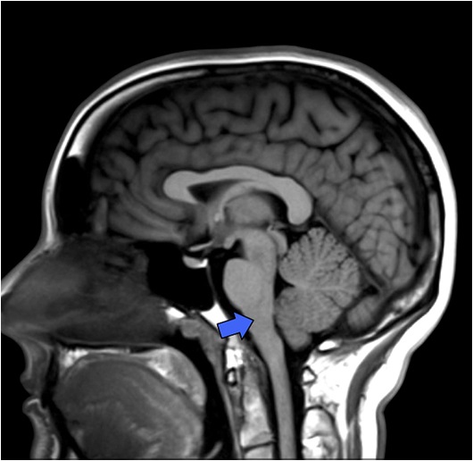
Identify the marked structure in the image:
| A. |
Midbrain |
| B. | Pons |
| C. | Medulla oblongata |
| D. |
Spinal cord |


|
Correct Answer � B Explanation |
 |

The marked structure in the image is the Pons. The pons is located just below the midbrain and above the medulla oblongata. It is visible on an MRI or CT scan as a larger bulge in the brainstem. In a sagittal section, the pons may appear as a larger, oval-shaped structure located below the midbrain and above the medulla oblongata.
Reasons to rule out other options
Option A – Midbrain: The midbrain is located in the uppermost part of the brainstem, just below the thalamus and above the pons. It is visible on an MRI or CT scan as a “bulge” in the brainstem. In a sagittal section, the midbrain may appear as a small round structure located above the pons and below the thalamus.
It is located anterior to the fourth ventricle and has a distinctive “butterfly” shape on axial views.
Option C – Medulla oblongata: The medulla oblongata is located at the bottom of the brainstem, just above the spinal cord. It is visible on an MRI or CT scan as a tapering structure that connects the brainstem to the spinal cord. In a sagittal section, the medulla oblongata may appear as a smaller, pointed structure located below the pons.
The medulla oblongata appears as a tapering structure that connects the brainstem to the spinal cord and is located at the bottom of the brainstem.
Option D – Spinal cord: The spinal cord is located within the spinal canal, which is formed by the vertebrae of the spine. It is not directly visible on an MRI or CT scan of the skull, but it can be visualized on an image of the spine. In a sagittal section, the spinal cord may appear as a central canal surrounded by white matter and gray matter.
The spinal cord is located within the spinal canal, which is formed by the vertebrae of the spine.