

MUSCULAR COMPARTMENT OF LEG
| A | Pectoralis major | |
| B | Extensor digitorum | |
| C | Flexor digitorum profundus | |
| D | Brachialis |
Which is an unlocker of knee?
| A | Popliteus | |
| B | biceps femoris | |
| C |
Sartorius |
|
| D |
Gastrocnemius |
Tibialis posterior originates from
| A | Tibia | |
| B |
Fibula |
|
| C |
Interosseus membrane |
|
| D |
All the above |
The muscle that acts primarily as an evertor of ankle inserted into the medial cuneiform is which of the following?
| A |
Peroneus longus |
|
| B |
Peroneus brevis |
|
| C |
Tibialis anterior |
|
| D |
Tibialis posterior |
Which of the following muscles is used in normal walk during stance and swing?
| A |
Popliteus |
|
| B |
Gastrocnemius |
|
| C |
Tibialis anterior |
|
| D |
Iliopsoas |
Among the following, the structure that passes deep to the flexor retinaculum is?
| A |
Tibialis anterior |
|
| B |
Tibialis posterior |
|
| C |
Peroneus brevis |
|
| D |
Peroneus longus |
Boundaries of Popliteal fossa
| A |
Biceps femoris |
|
| B |
Both heads of Gastrocnemius |
|
| C |
Adductor magnus |
|
| D |
All |
Violent inversion of the foot will lead to avulsion of tendon of which the following muscle attached to the tuberosity of the 5th metatarsal?
| A |
Peroneus brevis |
|
| B |
Peroneus longus |
|
| C |
Peroneus tertius |
|
| D |
Extensor digitorum brev |
Muscle acting both at knee and ankle joint is/are:
| A |
Gastrocnemius |
|
| B |
Soleus |
|
| C |
Plantaris |
|
| D |
A and c both |
Muscle(s) causing dorsiflexion of foot is/are:
| A |
Extensor digitorum longus |
|
| B |
Extensor hallucis longus |
|
| C |
Tibialis anterior |
|
| D |
All |
Planter flaxion is brought about by which of these muscles:
| A |
Plantaris |
|
| B |
FHL |
|
| C |
Soleus |
|
| D |
All |
Anterior compartment of leg contains all muscle except :
| A |
Peroneus brevis |
|
| B |
Peroneus tertius |
|
| C |
Extensor hallucis longus |
|
| D |
Tibialis anterior |
Identify the Muscle Marked as A in the Image


| A |
Sartorius |
|
| B |
Gastrocnemius |
|
| C |
Soleus |
|
| D |
Tibialis Posterior |
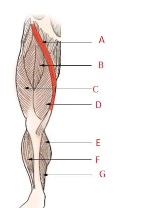
Identify the Muscle Marked as A in the Image
| A |
Sartorius |
|
| B |
Gastrocnemius |
|
| C |
Soleus |
|
| D |
Tibialis Posterior |
True about popliteus are all except‑
| A |
Flexor of knee |
|
| B |
Intracapsular origin |
|
| C |
Supplied by tibial nerve |
|
| D |
Causes locking of knee |
Which of the following dorsiflexes the foot –
| A |
Tibialis posterior |
|
| B |
Tibialis anterior |
|
| C |
Peroneus brevis |
|
| D |
Extensor digitorum brevis |
Action of tibialis anterior ‑
| A |
Plantar flexion of foot |
|
| B |
Adduction of foot |
|
| C |
Inversion of foot |
|
| D |
None of the above |
Action of popliteus muscle ‑
| A |
Medial rotation of femur |
|
| B |
Lateral rotation of femur |
|
| C |
Locking of knee |
|
| D |
Extension of knee |
Which is called as peripheral heart ‑
| A |
Popliteus |
|
| B |
Soleus |
|
| C |
Plantaris |
|
| D |
None |
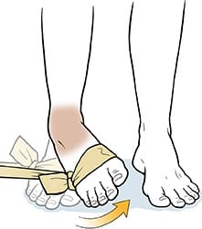
Action (Arrow) shown in photograph is due to action of

| A |
Tibialis anterior |
|
| B |
Tibialis posterior |
|
| C |
Soleus |
|
| D |
Peroneus longus |
| A | Pectoralis major | |
| B | Extensor digitorum | |
| C | Flexor digitorum profundus | |
| D | Brachialis |
Extensor digitorum
Hybrid muscles are the muscles having more than one nerve supply
Pctoralis major: lateral & medial pectoral nerves
Brachialis: additional proprioceptive fibres from radial nerve, in addition to its muscular nerve, musculocutaneous nerve.
Extensor digitorum is supplied by the posterior interosseous nerve only.
Which is an unlocker of knee?
| A |
Popliteus |
|
| B |
biceps femoris |
|
| C |
Sartorius |
|
| D |
Gastrocnemius |
Popliteus
- Locking or screw home mechanism: Conjunct medial rotation of the femur on the tibia in later stages of extension due to quadriceps femoris locks the knee joint.
- Unlocking of the knee joint: At beginning of flexion from the full extension with foot flexed, lateral rotation by Popliteus muscle unlocks the joint.
Tibialis posterior originates from
| A | Tibia | |
| B | Fibula | |
| C |
Interosseus membrane |
|
| D |
All the above |
ORIGIN:
- The tibialis posterior muscle originates on the inner posterior borders of the tibia and fibula.
- It is also attached to the interosseous membrane, which attaches to the tibia and fibula.
INSERTION:
- The tendon of the tibialis posterior muscle (sometimes called the posterior tibial tendon) descends posterior to the medial malleolus and terminates by dividing into plantar, main, and recurrent components.
- The main portion inserts into the tuberosity of the navicular and the plantar surface of the medial cuneiform.
- The plantar portion inserts into the bases of the second, third and fourth metatarsals, the intermediate and lateral cuneiforms and the cuboid.
- The recurrent portion inserts into the sustentaculum tali of the calcaneus.
| A |
Peroneus longus |
|
| B |
Peroneus brevis |
|
| C |
Tibialis anterior |
|
| D |
Tibialis posterior |
The peroneus longus and brevis act primarily as evertors, with the peroneus brevis being the stronger of the two. In the question, the evertor inserted to the medial cuneiform, which is peroneus longus.
The peroneus brevis inserts on the base of the fifth metatarsal and the peroneus longus courses under the cuboid to insert on the base of the first metatarsal and medial cuneiform.
The tibialis anterior is the dorsiflexor of foot at the ankle joint. It is the invertor of the foot at the midtarsal and subtalar joints.
Tibialis posterior is the principal invertor of the foot.
Which of the following muscles is used in normal walk during stance and swing?
| A |
Popliteus |
|
| B |
Gastrocnemius |
|
| C |
Tibialis anterior |
|
| D |
Iliopsoas |
Normal walking constitutes of the stance phase (when at least one leg is in contact with the ground) and swing phase (when the leg is in the air).
| A |
Tibialis anterior |
|
| B |
Tibialis posterior |
|
| C |
Peroneus brevis |
|
| D |
Peroneus longus |
Tibialis posterior is the deepest and most centrally located muscle in the deep posterior compartment of the leg. It arises from the upper posterior aspect of tibia, fibula and interosseous membrane and passes medially deep to the flexor retinaculum. Its tendons flares and inserts into the navicular, tarsal bones, and bases of the second, to fourth metatarsals.
- Tibialis posterior tendon
- Flexor digitorum longus tendon
- Flexor hallucis longus tendon
- Posterior tibial artery
- Tibial nerve.
| A |
Biceps femoris |
|
| B |
Both heads of Gastrocnemius |
|
| C |
Adductor magnus |
|
| D |
All |
A. i.e. Biceps femoris; B. i.e. Both heads of gastrocnemius; C. i.e. Adductor magnus
Violent inversion of the foot will lead to avulsion of tendon of which the following muscle attached to the tuberosity of the 5th metatarsal?
| A |
Peroneus brevis |
|
| B |
Peroneus longus |
|
| C |
Peroneus tertius |
|
| D |
Extensor digitorum brev |
A i.e. Peroneus brevis
Violent inversion of foot may cause avulsion of tuberosity of 5n1, metatarsal base, the insertion of peroneus (fibularis) brevis muscleQ.
| A |
Gastrocnemius |
|
| B |
Soleus |
|
| C |
Plantaris |
|
| D |
A and c both |
A i.e. Gastrocnemius C i.e. Plantaris
Muscle(s) causing dorsiflexion of foot is/are:
| A |
Extensor digitorum longus |
|
| B |
Extensor hallucis longus |
|
| C |
Tibialis anterior |
|
| D |
All |
A i.e. Extensor digitorum longus; B i.e. Extensor hallucis longus; C i.e. Tibialis anterior
Planter flaxion is brought about by which of these muscles:
| A |
Plantaris |
|
| B |
FHL |
|
| C |
Soleus |
|
| D |
All |
A. i.e. Plantans; B. i.e. FHL; C. i.e. Soleus
Anterior compartment of leg contains all muscle except :
| A |
Peroneus brevis |
|
| B |
Peroneus tertius |
|
| C |
Extensor hallucis longus |
|
| D |
Tibialis anterior |
A. i.e. Peroneus brevis
Identify the Muscle Marked as A in the Image
| A |
Sartorius |
|
| B |
Gastrocnemius |
|
| C |
Soleus |
|
| D |
Tibialis Posterior |
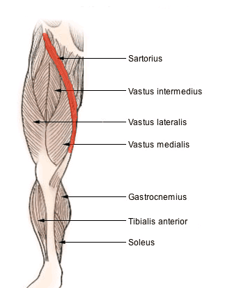
Identify the Muscle Marked as A in the Image
| A |
Sartorius |
|
| B |
Gastrocnemius |
|
| C |
Soleus |
|
| D |
Tibialis Posterior |

True about popliteus are all except‑
| A |
Flexor of knee |
|
| B |
Intracapsular origin |
|
| C |
Supplied by tibial nerve |
|
| D |
Causes locking of knee |
Popliteus
Popliteus is a deep muscle of posterior compartment of leg.
Features of popletius are –
Origin
- Lateral surface of lateral condyle of femur, origin is intracapsular.
- Outer margin of lateral meniscus of knee.
Insertion
- Posterior surface of shaft of tibia above soleal line.
Nerve supply
- Tibial nerve
Action
- Ulocks knee joint by lateral rotation of femur on tibia prior flexion.
- Accessory flexor of knee.
| A | Tibialis posterior | |
| B |
Tibialis anterior |
|
| C |
Peroneus brevis |
|
| D |
Extensor digitorum brevis |
Tibialis anterior
Action of tibialis anterior ‑
| A |
Plantar flexion of foot |
|
| B |
Adduction of foot |
|
| C |
Inversion of foot |
|
| D |
None of the above |
Ans. is ‘c’ i.e., Inversion of foot
Action of popliteus muscle ‑
| A |
Medial rotation of femur |
|
| B |
Lateral rotation of femur |
|
| C |
Locking of knee |
|
| D |
Extension of knee |
Ans. is ‘b’ i.e., Lateral rotation of femur
Ponliteus
* Popliteus is a deep muscle of posterior compartment of leg.
* Features of popletius are ‑
Origin
* Lateral surface of lateral condyle of femur, origin is intracapsular.
* Outer margin of lateral meniscus of knee.
Insertion
* Posterior surface of shaft of tibia above soleal line.
Nerve supply
* Tibial nerve
Action
* Ulocks knee joint by lateral rotation of femur on tibia prior flexion.
* Accessory flexor of knee
| A | Popliteus | |
| B |
Soleus |
|
| C |
Plantaris |
|
| D |
None |
Ans. is ‘b’ i.e., Soleus
Soleus is known as ‘peripheral heart’ as it helps in venous return from lower limb.
Action (Arrow) shown in photograph is due to action of
| A |
Tibialis anterior |
|
| B |
Tibialis posterior |
|
| C |
Soleus |
|
| D |
Peroneus longus |
Ans:A. Tibialis Anterior.
Inversion of foot is shown in the image.

