

Penetrating injury
| A | More than 2 cm deep injury | |
| B |
Injury to any internal neck organ |
|
| C |
Violation of platysma |
|
| D |
Sharp instrument |
True about esophageal injury:
| A |
Most common after penetrating injury |
|
| B |
Can lead to mediastinal collection |
|
| C |
Barium swallow is diagnostic |
|
| D |
All |
Commonest cause of death in penetrating injury of chest –
| A |
Tracheobronchial injury |
|
| B |
Oesophageal rupture |
|
| C |
Pulmonary laceration |
|
| D |
Chylothorax |
Sympathetic ophthalmitis results due to:
| A |
Penetrating injury of ciliary body |
|
| B |
Uveitis |
|
| C |
Glaucoma |
|
| D |
Trachoma |
Most common organ injured in penetrating injury of the abdomen:
| A |
Liver |
|
| B |
Spleen |
|
| C |
Small bowel |
|
| D |
None of these |
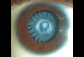
A patient presented with a penetrating injury of metallic foreign body.The condition of the eye is shown in the image.Which is the most probable metal in the foreign body?

| A |
Iron. |
|
| B |
Copper. |
|
| C |
Lead. |
|
| D |
Mercury. |
A patient presented with a penetrating injury of metallic foreign body. The condition of the eye is shown in the picture below.In this condition the electrolytically dissociated ions of metal are ?


| A |
Deposited under the membraneous structures of the eye. |
|
| B |
Combine with intracellular proteins and produce degenerative changes. |
|
| C |
Produce irritative reaction at the local site. |
|
| D |
All of the above. |
Penetrating injury to NECK is defined as
| A |
More than 2 cm deep injury |
|
| B |
Injury to any internal neck organ |
|
| C |
Violation of platysma |
|
| D |
Sharp instrument |
C i.e. Violating platysma
Penetrating neck trauma involves a gun shot or sharp object (stab, puncture wound & impalement injuries) penetrating the skin and volating the platysma layer of neckQ
| A |
Most common after penetrating injury |
|
| B |
Can lead to mediastinal collection |
|
| C |
Barium swallow is diagnostic |
|
| D |
All |
Ans is All
- Most injuries to the esophagus are from penetrating trauma. Associated injuries are the rule.
- Symptoms include odynophagia (pain on swallowing foods or fluids), subcutaneous or mediastinal emphysema, pleural effusion, air in the retro-esophageal space and unexplained fever within 24 hours of injury. Mediastinal and deep cervical emphysema must be seen as evidence of an aerodigestive injury until proven otherwise.
- If the diagnosis is made early, primary surgical repair is generally possible. If the diagnosis is delayed for more than 24 hours, primary repair may be impractical, with diversion and drainage being the only alternative. The major morbidity and mortality associated with esophageal injuries are the result of delay in diagnosis, which underscores the necessity to aggressively exclude esophageal injury during the initial evaluation of a patient with neck injury. “The mortality rate rises exponentially if treatment is delayed for more than 12-24 hours.”- Bailey and Love
- Esophagoscopy can be performed to evaluate the esophagus, but injuries have been missed with this technique alone. Therefore patients at risk should also undergo a soluble contrast esophagram looking for extravasation of contrast. If no extravasation is seen, a barium esophagram should be performed for greater detail. Failure to identify esophageal injuries leads to fulminant mediastinitis that is often fatal.
- Treatment consists of early debridement, primary repair, and drainage if identified within 24 hours after injury. Injuries diagnosed after 24 hours with mediastinal contamination are treated by cervical esophagostomy and distal feeding access. Esophageal resection is rarely needed but may be indicated in patients with esophageal necrosis or severe mediastinitis.. A drain should be left in place after all esophageal repairs.
Commonest cause of death in penetrating injury of chest –
| A | Tracheobronchial injury | |
| B |
Oesophageal rupture |
|
| C |
Pulmonary laceration |
|
| D |
Chylothorax |
Ans is ‘a’ ie Tracheobronchial injury
- Tracheobronchial injuries are so fatal that most pts. die at the scene or during transport as a result of poor ventilation.
- The other options are so easy to rule out as they are rarely the cause of death.
- I could not find the MC cause of death after penetrating injury but among the 4 options provided Tracheobronchial is definitely the right answer. No doubts about it.
| A |
Penetrating injury of ciliary body |
|
| B |
Uveitis |
|
| C |
Glaucoma |
|
| D |
Trachoma |
Ans. A: Penetrating Injury of Ciliary Body
Sympathetic ophthalmitis or ophthalmia almost always results from a penetrating wound. others
Incarceration of the iris or lens capsule is more likely to set up sympathetic ophthalmitis than oers
Sympathetic ophthalmia/ SO
- It is a granulomatous uveitis of both eyes following trauma to one eye.
- It can leave the patient completely blind.
- Symptoms may develop from days to several years after a penetrating eye injury.
- Floating spots and loss of accommodation are among the earliest symptoms.
- The disease may progress to severe iridocyclitis with pain and photophobia.
- Commonly the eye remains relatively painless while the inflammatory may occur. disease spreads through the uvea, where characteristic focal infiltrates in the choroid named Dalen-Fuchs nodules can be seen.
- Papilledema, secondary glaucoma, vitiligo, and poliosis of the eyelashes may accompany SO.
- In approximately 80% of cases, the uveitis appears within 2-12 weeks after injury, and 90% occur within 1 year from the time of injury.
- Sympathetic ophthalmia is currently thought to be an autoimmune inflammatory response toward ocular antigens, specifically a delayed hypersensitivity to melanin-containing structures from the outer segments of the photoreceptor layer of the retina.
- The immune system, which normally is not exposed to ocular antigens, is introduced to the contents of the eye following traumatic injury.
- Once exposed, it senses these antigens as foreign, and begins attacking them.
- The onset of this process can be from days to years after the inciting traumatic event.
Diagnosis
- Diagnosis is clinical, seeking a history of eye injury.
- An important differential diagnosis is Vogt-Koyanagi-Harada syndrome (VKH), which is thought to have the same pathogenesis, without a history of surgery or penetrating eye injury.
Prevention and treatment
Definitive prevention of SO requires prompt (within the first 7 to 10 days following injury) enucleation of the injured eye.
Evisceration—the removal of the contents of the globe while leaving the sclera and extraocular muscles intact—is easier to perform, offers long-term orbital stability, and is more aesthetically pleasing.
- There is concern, however, that evisceration may lead to a higher incidence of SO compared to enucleation
- The first choice of treatment may not be enucleation or evisceration, especially if there is a chance that the injured eye may regain some function
- Immunosuppressive therapy is the mainstay of treatment for SO.
- Mild cases may be treated with local application of corticosteroids and pupillary dilators.
- More severe or progressive cases require high-dose systemic corticosteroids for months to years.
Patients who become resistant to corticosteroids or develop side effects of long-term corticosteroid therapy (osteoporosis and pathologic fractures, mental status changes, etc.), may be candidates for therapy with chlorambucil, cyclophosphamide, or ciclosporin.
| A |
Liver |
|
| B |
Spleen |
|
| C |
Small bowel |
|
| D |
None of these |
Ans- C- Small Bowel
- The most common cause is a stab or gunshot.
- The most common organs injured are the small bowel (50%), large bowel (40%), liver (30%), and intra-abdominal vascular (25%).
- When the injury is close range, there is more kinetic energy than those injuries sustained from a distance. Even though most gunshot wounds typically have a linear projection, the high-energy wounds are associated with unpredictable injuries.
- There may also be secondary missile injuries from bone or bullet fragments. Stab wounds that penetrate the abdominal wall are difficult to assess.
Pathophysiology
- As a projectile passes through tissue, it decelerates and transfers kinetic energy to the tissue. Increased velocity causes more damage than mass. Kinetic energy increases with the square of the velocity.
- The space left by tissue that is destroyed by the penetrating object forms a cavity, and this is called permanent cavitation. In addition to damage to the tissues they contact, medium- and high-velocity projectiles result in a secondary cavitation injury as the object enters the body, it creates a pressure wave forcing tissue out of the way, creating a cavity. The tissues move back into place, eliminating the cavity, but the cavitation has already done considerable damage.
- The characteristics of the damaged tissue determine the severity of the injury: the denser the tissue, the greater the amount of energy transmitted to it.
| A |
Iron. |
|
| B |
Copper. |
|
| C |
Lead. |
|
| D |
Mercury. |
Ans:B.)Copper.
The condition shown in the picture above represents Chalcosis (Sunflower Catacract and Kayser-Fleischer ring)
- It is defined as tissue damage to ocular structures as a direct result of retained metal particles following penetrating globe injury by an Intraocular Foreign Body(IOFB)
- Metallosis can occur as a consequence of numerous types of metals, but iron and copper alloys are the two most common.
- When an IOFB contains iron, the form of metallosis that develops is referred to as siderosis.
- Copper-containing IOFBs with less than 85% copper content cause chalcosis.
| Findings | Siderosis | Chalcosis |
| Ocular Anatomical Features | ||
| Cornea | Usually normal, but the stroma may develop a diffuse brown haze late in the clinical course | Kayser-Fleischer ring |
| Iris | Heterochromia with the affected side having a brown discoloration |
Heterochromia with the affected side having a greenish color |
| Lens | Diffuse, brownish discoloration of the anterior capsule and generalized yellowing of cortex | Classic sunflower cataract of the anterior capsule |
| Retina | RPE degeneration affecting peripheral fundus first and posterior segment later | Refractile deposits in the macular region,with sparing of the periphery |
| Vitreous | Brownish opacification | Copperlike opacification |
| Magnetic Properties of IOFB | Yes | No |
| Pathophysiology | Iron ions deposited intracellularly | Copper ions deposited in basement membranes |
| Clinical course if IOFB is not removed | Slow, relentless progression with loss of all vision | Variable, but may preserve reasonably good visual function |
| A |
Deposited under the membraneous structures of the eye. |
|
| B |
Combine with intracellular proteins and produce degenerative changes. |
|
| C |
Produce irritative reaction at the local site. |
|
| D |
All of the above. |
Ans:A.)Deposited under the membraneous structures of the eye.
The condition shown in the picture above represents Chalcosis (Sunflower Catacract and Kayser-Fleischer ring)
Some patient’s with Wilson’s disease will have copper deposition (chalcosis) in the lens. This is termed a “sunflower” cataract because of its appearance when fully developed. The copper deposits are in and beneath the lens capsule, usually on the anterior surface.
In chalcosis bulbi the electrolytically dissociated ions of copper are deposited under the membraneous structures of the eye.
Chalcosis
- It refers to the specific changes produced by the alloy of copper in the eye.
- Copper from the alloy are dissociated electrolytically and are deposited under the membranous structure of the eye.
- Clinical manifestations of chalcosis includes:
- Kayser Fleischer rings: due to deposition of copper under peripheral parts of Descement’s membrane.
- Sunflower cataract: due to deposition of copper under the posterior capsule of the lens.
- Deposition of golden plaques at the posterior pole of retina which reflects the light with a metallic sheen.