
FORAMENS OF SKULL
| A | Maxillary artery | |
| B |
Mandibular nerve |
|
| C |
Middle meningeal artery |
|
| D |
Spinal accessory nerve |
Structure passing through foramen Rotundum?
| A |
Maxillary artery |
|
| B |
Maxillary nerve |
|
| C |
Middle meningeal artery |
|
| D |
Spinal accessory nerve |
| A | Greater wing of sphenoid and lesser wing of sphenoid. | |
| B | Greater wing of sphenoid and palatine process of maxilla. | |
| C | Lesser wing of sphenoid and palatine process of maxilla. | |
| D | Greater wing and lesser wing of sphenoid and body of sphenoid. |
Foramen spinosum transmits the following?
| A |
Meningeal branch of the mandibular nerve, middle meningeal artery |
|
| B |
Emissary veins from the cavernous sinus |
|
| C |
Both A and B |
|
| D |
None of the above |
Which of the following opening in the base of the skull transmits the third branch of trigeminal nerve?
| A |
Foramen ovale |
|
| B |
Foramen lacerum |
|
| C |
Foramen magnum |
|
| D |
Foramen spinosum |
Which of the following cranial nerve travels through the jugular foramen in the base of the skull?
| A |
3rd branch of trigeminal nerve |
|
| B |
Abducens nerve |
|
| C |
Facial nerve |
|
| D |
Glossopharyngeal nerve |
Middle meningeal artery courses along which of the following opening in the base of the skull?
| A |
Foramen ovale |
|
| B |
Foramen lacerum |
|
| C |
Foramen spinosum |
|
| D |
Foramen rotundum |
Which of the following structure is passing through foramen rotundum?
| A |
Maxillary artery |
|
| B |
Maxillary nerve |
|
| C |
Middle meningeal artery |
|
| D |
Spinal accessory nerve |
True about relation of epiploic foramen is :
| A |
Portal vein posteriorly |
|
| B |
IVC inferiorly |
|
| C |
Hepatic art superiorly |
|
| D |
Bile duct anteriorly |
Primary and secondary palates are divided by
| A |
Greater palatine foramen |
|
| B |
Canine teeth |
|
| C |
Alveolar arch |
|
| D |
Incisive foramen |
Mandibular nerve passes through following foramen:
| A |
F. ovale |
|
| B |
F. rotundum |
|
| C |
F. spinosum |
|
| D |
F. lacerum |
All of the following nerves pass through Jugular foramen except:
| A |
9th |
|
| B |
10th |
|
| C |
11th |
|
| D |
12th |
Structures passing through superior orbital fissure:
| A |
Cranial nerve VI |
|
| B |
Cranial nerve I |
|
| C |
Cranial nerve II |
|
| D |
Ophthalmic nerve |
Structures passing through superior orbital fissure:
| A |
Oculomotor nerve |
|
| B |
Trochlear nerve |
|
| C |
Lacrimal nerve |
|
| D |
All |
Structure passing through the foramen manum are
| A |
Spinal cord |
|
| B |
Vertebral artery |
|
| C |
Internal jugular vein |
|
| D |
All |
Structures passing through foramen magnum include all
| A |
Spinal accessory nerve |
|
| B |
Spinal cord |
|
| C |
Vertebral artery |
|
| D |
Vertebral venous plexus |
True about foramen of Morgagni:
| A |
It is femoral canal |
|
| B |
It is a diaphragmatic opening |
|
| C |
Superior epigastric vessels passes through it |
|
| D |
B and C |
Which of the following regarding mandibular nerve is correct-
| A |
Branch of facial nerve |
|
| B |
Purely motor |
|
| C |
Passes through foramen ovale |
|
| D |
Related to sphenopalatine ganglion |
Hernia through foramen of Bochdalek true is –
| A |
Congeniatal hernia |
|
| B |
Is asymptomatic |
|
| C |
Seen especially in males |
|
| D |
Least common |
Trochlear and abducent nerve pass through
| A |
Optic canal |
|
| B |
Superior orbital fissure |
|
| C |
Inferior orbital fissure |
|
| D |
Infraorbital foramen |
Structures Passing Through The Foramen Marked in the Diagram are all EXCEPT?

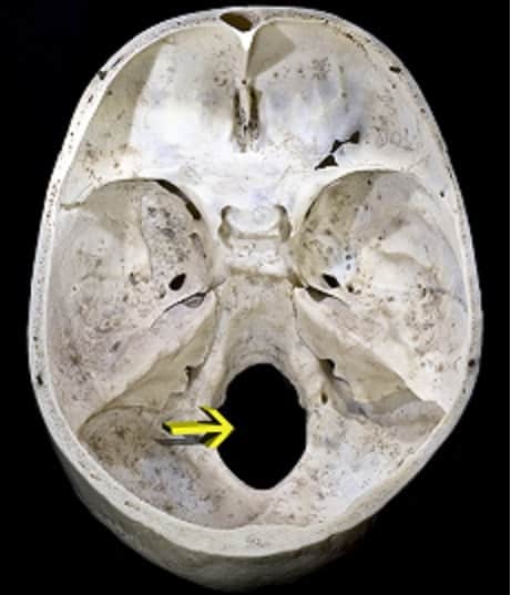
| A |
Spinal accessory nerve |
|
| B |
Spinal cord |
|
| C |
Vertebral artery |
|
| D |
Vertebral venous plexus |
Identify the Foramen marked in the Picture


| A |
Incisive Foramen |
|
| B |
Mental Foramen |
|
| C |
Greater Palatine foramina |
|
| D |
Lesser Palatine foramina |
Optic canal is a part of ‑
| A |
Lesser wing of sphenoid |
|
| B |
Greater wing of sphenoid |
|
| C |
Ethmoid |
|
| D |
Pterygoid |
Mandibular nerve passes through ‑
| A |
Formanen rotundum |
|
| B |
Foramen lacerum |
|
| C |
Stylomastoid foramen |
|
| D |
Foramen ovale |
All are true about boundaries of epiploic foramen except –
| A |
Anterior: portal vein & hepatic artery |
|
| B |
Posterior: IVC |
|
| C |
Inferior: liver |
|
| D |
Inferior: duodenum |
Which of the following is wrong regarding ophthalmic artery ‑
| A |
Present in dura along with optic nerve |
|
| B |
Supplies anterior ethmoidal sinus |
|
| C |
Artery to retina is end artery |
|
| D |
Leaves orbit through inferior orbital fissure |
True about vertebral artery ‑
| A |
A branch of thyrocervical trunk |
|
| B |
Enters skull through foramen magnum |
|
| C |
Unite to form posterior cerebral artery |
|
| D |
A small artery |
Boundries of foramen of Winslow is formed by all except ‑
| A |
IVC |
|
| B |
Liver |
|
| C |
2nd part of duodenum |
|
| D |
Suprarenal gland |
Which of the following is incorrectly matched ‑
| A |
Foramen rotundum – Maxillary nerve |
|
| B |
Foramen ovale – Mandibular nerve |
|
| C |
Foramen spinesum – Middle meningeal artery |
|
| D |
Jugular foramen – External jugular vein |
Superior orbital fissure syndrome – following nerves are affected ‑
| A |
C.N. 1, 2, 4, 6 |
|
| B |
C.N. 2, 3, 4, 6 |
|
| C |
C.N. 3, 4, 5, 6 |
|
| D |
C.N. 1, 2, 3, 4, 5 |
Structures Passing Through The Foramen Marked in the Diagram are all EXCEPT?

| A |
Spinal accessory nerve |
|
| B |
Spinal cord |
|
| C |
Vertebral artery |
|
| D |
Vertebral venous plexus |
This arrow showing Foramen Spinosum,,Contents are ?

| A |
Vertebral venous plexus |
|
| B |
Vertebral artery |
|
| C |
Middle meningeal artery |
|
| D |
Maxillary nerve |
The canal marked by an arrow, is a part of?


| A |
Lesser wing of sphenoid |
|
| B |
Greater wing of sphenoid |
|
| C |
Ethmoid |
|
| D |
Pterygoid |
Contents of jugular foramen are?


| A |
Glossopharyngeal nerve |
|
| B |
vagus nerve |
|
| C |
Accessory nerve |
|
| D |
All the above |
Structure passing through both greater and lesser sciatic foramen is ‑
| A |
Pudendal nerve |
|
| B |
Sciatic nerve |
|
| C |
Superior gluteal nerve |
|
| D |
Inferior gluteal nerve |
All pass through jugular foramen except
| A |
Emissary vein |
|
| B |
Vagus nerve |
|
| C |
Mandibular nerve |
|
| D |
Internal jugular vein |
Foramen ovale transmits all except
| A |
Emissary vein |
|
| B |
Mandibular nerve |
|
| C |
Lesser petrosal nerve |
|
| D |
Middle meningeal artery |
Structure passing through Foramen Ovale is?
| A |
Maxillary artery |
|
| B |
Mandibular nerve |
|
| C |
Middle meningeal artery |
|
| D |
Spinal accessory nerve |
Mandibular nerve
Structure passing through foramen Rotundum?
| A |
Maxillary artery |
|
| B |
Maxillary nerve |
|
| C |
Middle meningeal artery |
|
| D |
Spinal accessory nerve |
Maxillary nerve
| A | Greater wing of sphenoid and lesser wing of sphenoid. | |
| B | Greater wing of sphenoid and palatine process of maxilla. | |
| C | Lesser wing of sphenoid and palatine process of maxilla. | |
| D | Greater wing and lesser wing of sphenoid and body of sphenoid. |
Foramen rotundum: It is situated behind the medial end of superior orbital fissure. It perforates the greater wing of sphenoid and transmits the maxillary nerve from the trigeminal ganglion to the pterygopalatine fossa.
Foramen ovale: It lies posterolateral to the foramen rotundum. It perforates the greater wing of sphenoid and transmits the large sensory root and small motor root of the mandibular nerve to the infratemporal fossa.
Ref: Tsai L.M., Kamenetzky S.A. (2010). Chapter 37. The Eye & Ocular Adnexa. In G.M. Doherty (Ed), CURRENT Diagnosis & Treatment: Surgery, 13e.
Foramen spinosum transmits the following?
| A |
Meningeal branch of the mandibular nerve, middle meningeal artery |
|
| B |
Emissary veins from the cavernous sinus |
|
| C |
Both A and B |
|
| D |
None of the above |
- Foramen ovale
- Foramen lacerum
- Foramen spinosum
- Foramen rotundum
- Superior orbital fissure
|
Foramina in middle cranial fossa |
Location |
Content |
|
Foramen ovale |
Posterolateral to the foramen rotundum |
Accessory meningeal artery Mandibular nerve (V3) Lesser petrosal nerve (occasionally) accessory meningeal vein |
|
Foramen spinosum |
Posterolateral to the foramen ovale |
Middle meningeal artery and vein Meningeal branch of the mandibular nerve (V3) Sympathetic plexus |
|
Foramen lacerum |
Lies superomedially to the foramen spinosum |
Internal carotid artery, Artery of pterygoid canal Nerve of pterygoid canal |
|
Superior orbital fissure |
Slit like opening between the lesser and greater wings of the sphenoid |
Oculomotor nerve (III) Trochlear nerve (IV) Lacrimal, frontal and nasociliary branches of Ophthalmic nerve (V1) Abducent nerve (VI) Orbital branch of middle meningeal artery Recurrent branch of lacrimal artery Superior orbital vein Superior ophthalmic vein |
|
Foramen rotundum |
Below and behind the medial end of the superior orbital fissure |
Maxillary nerve (V2) |
| A | Foramen ovale | |
| B |
Foramen lacerum |
|
| C |
Foramen magnum |
|
| D |
Foramen spinosum |
- Foramen lacerum transmits the internal carotid artery.
- Foramen magnum transmits the medulla and its membranes, the spinal accessory nerves, the vertebral arteries, and the anterior and posterior spinal arteries.
Which of the following cranial nerve travels through the jugular foramen in the base of the skull?
| A |
3rd branch of trigeminal nerve |
|
| B |
Abducens nerve |
|
| C |
Facial nerve |
|
| D |
Glossopharyngeal nerve |
Middle meningeal artery courses along which of the following opening in the base of the skull?
| A |
Foramen ovale |
|
| B |
Foramen lacerum |
|
| C |
Foramen spinosum |
|
| D |
Foramen rotundum |
|
Foramen |
Structures |
|
Cribriform plate of ethmoid |
Olfactory nerves |
|
Optic foramen |
Optic nerve, ophthalmic artery, meninges |
|
Superior orbital fissure |
Oculomotor, trochlear, and abducens nerves; ophthalmic division of trigeminal nerve; superior ophthalmic vein |
|
Foramen rotundum |
Maxillary division of trigeminal nerve, small artery and vein |
|
Foramen ovale |
Mandibular division of trigeminal nerve, vein |
|
Foramen lacerum |
Internal carotid artery, sympathetic plexus |
|
Foramen spinosum |
Middle meningeal artery and vein, meningeal branch of mandibular nerve |
|
Internal acoustic meatus |
Facial and vestibulocochlear nerves, internal auditory artery |
|
Jugular foramen |
Glossopharyngeal, vagus, and spinal accessory nerves; sigmoid sinus |
|
Hypoglossal canal |
Hypoglossal nerve |
|
Foramen magnum |
Medulla and meninges, spinal accessory nerve, vertebral arteries, anterior and posterior spinal arteries |
| A | Maxillary artery | |
| B |
Maxillary nerve |
|
| C |
Middle meningeal artery |
|
| D |
Spinal accessory nerve |
True about relation of epiploic foramen is :
| A |
Portal vein posteriorly |
|
| B |
IVC inferiorly |
|
| C |
Hepatic art superiorly |
|
| D |
Bile duct anteriorly |
D. i.e. Bile duct anteriorly
Primary and secondary palates are divided by
| A |
Greater palatine foramen |
|
| B |
Canine teeth |
|
| C |
Alveolar arch |
|
| D |
Incisive foramen |
D. i.e. Incisive foramen
The incisive foramen is dividing landmark between the primary & secondary palateQ; and anterior & posterior cleft deformities
Mandibular nerve passes through following foramen:
| A |
F. ovale |
|
| B |
F. rotundum |
|
| C |
F. spinosum |
|
| D |
F. lacerum |
A i.e. Foramen ovale
Vidian nerve and artery pass through pterygoid canal.
Foramen spinosum passes MEN i.e. Middle meningeal artery, Emissary vein & Nervous spinosus (meningeal br. of mandibular nerve).
Foramen ovale passes MALE i.e. Mandibular nerve, Accessory meningeal artery, Lesser petrosal nerve and Emissary vein.
| A | 9th | |
| B |
10th |
|
| C |
11th |
|
| D |
12th |
D i.e. 12th nerve
Structures passing through superior orbital fissure:
| A |
Cranial nerve VI |
|
| B |
Cranial nerve I |
|
| C |
Cranial nerve II |
|
| D |
Ophthalmic nerve |
A i.e. Cranial nerve VI
Structures passing through superior orbital fissure:
| A |
Oculomotor nerve |
|
| B |
Trochlear nerve |
|
| C |
Lacrimal nerve |
|
| D |
All |
A. i.e. Oculomotor nerve; B i.e. Trochlear nerve; C i.e. Lacrimal nerve
Structure passing through the foramen manum are
| A |
Spinal cord |
|
| B |
Vertebral artery |
|
| C |
Internal jugular vein |
|
| D |
All |
B i.e. Vertebral artery
Structures passing through foramen magnum include all
| A |
Spinal accessory nerve |
|
| B |
Spinal cord |
|
| C |
Vertebral artery |
|
| D |
Vertebral venous plexus |
B. i.e. Spinal cord
Lower part of medulla oblongata (not the spinal cord) passes through posterior part of foramen magnum, and vertebral arteries are transmitted through the subarachnoid space in foramen magnum.
| A |
It is femoral canal |
|
| B |
It is a diaphragmatic opening |
|
| C |
Superior epigastric vessels passes through it |
|
| D |
B and C |
B i.e. It is an opening through diaphragm C i.e. Superior epigastric vessels passes through it.
Which of the following regarding mandibular nerve is correct-
| A |
Branch of facial nerve |
|
| B |
Purely motor |
|
| C |
Passes through foramen ovale |
|
| D |
Related to sphenopalatine ganglion |
C i.e. Passes through foramen ovale
Hernia through foramen of Bochdalek true is –
| A |
Congeniatal hernia |
|
| B |
Is asymptomatic |
|
| C |
Seen especially in males |
|
| D |
Least common |
Answer is ‘a’ i.e. Congenital hernia
Trochlear and abducent nerve pass through
| A |
Optic canal |
|
| B |
Superior orbital fissure |
|
| C |
Inferior orbital fissure |
|
| D |
Infraorbital foramen |
Trochlear and Abducent nerves exit the cranium through the superior orbital fissure.
The optic nerve runs backward and medially and passes through the optic canal to enter the middle cranial fossa
Inferior orbital fissure transmits maxillary nerve, the zygomatic nerve, etc.
Infraorbital foramen transmits the infraorbital nerve and vessels
Structures Passing Through The Foramen Marked in the Diagram are all EXCEPT?
| A |
Spinal accessory nerve |
|
| B |
Spinal cord |
|
| C |
Vertebral artery |
|
| D |
Vertebral venous plexus |
The Structure Marked is Foramen Magnum
Lower part of medulla oblongata (not the spinal cord) passes through posterior part of foramen magnum, and vertebralarteries are transmitted through the subarachnoid space in foramen magnum
Apart from the transmission of the medulla oblongata and its membranes, the foramen magnum transmits the vertebral arteries, the anterior and posterior spinal arteries, the tectorial membranes and alar ligaments. It also transmits the accessory nerve into the skull.
Identify the Foramen marked in the Picture
| A |
Incisive Foramen |
|
| B |
Mental Foramen |
|
| C |
Greater Palatine foramina |
|
| D |
Lesser Palatine foramina |
Mental Foramen is marked in the Diagram.
- The mental foramen is one of two foramina (openings) located on the anterior surface of the mandible.
- It transmits the terminal branches of the inferior alveolar nerve and vessels (the mental artery).
- The mental foramen descends slightly in toothless individuals.
Optic canal is a part of ‑
| A |
Lesser wing of sphenoid |
|
| B |
Greater wing of sphenoid |
|
| C |
Ethmoid |
|
| D |
Pterygoid |
Ans. is ‘a’ i.e., Lesser wing of sphenoid
The optic nerve leaves the orbit is the optic canal to enter the cranial vault.
The optic canal is the most posterior landmark of the orbit. It measures 10 mm in length.
The thin piece of bone separating the optic canal from the superior orbital fissure is the optic strut.
The optic strut and optic canal are a part of the lesser wing of sphenoid bone.
| A | Formanen rotundum | |
| B |
Foramen lacerum |
|
| C |
Stylomastoid foramen |
|
| D |
Foramen ovale |
Foramen ovale
All are true about boundaries of epiploic foramen except –
| A |
Anterior: portal vein & hepatic artery |
|
| B |
Posterior: IVC |
|
| C |
Inferior: liver |
|
| D |
Inferior: duodenum |
Epiploic foramen (foramen of Winslow or aditus to lesser sac) is a slit-like opening through which lesser sac communicates with greater sac. It is situated at the level of the T12 vertebra. Its boundaries are:-
- Anterior:- Right free margin of lesser omentum (contains portal vein, hepatic artery proper and bile duct).
- Posterior:- IVC, right suprarenal gland and T12 vertebra.
- Superior:- Caudate lobe of the liver.
- Inferior:- 1st part of the duodenum and horizontal part of the hepatic artery.
| A |
Present in dura along with optic nerve |
|
| B |
Supplies anterior ethmoidal sinus |
|
| C |
Artery to retina is end artery |
|
| D |
Leaves orbit through inferior orbital fissure |
OPHTHALMIC ARTERY
Origin
The ophthalmic artery is a branch of the cerebral part of the internal carotid artery, given off medial to the anterior clinoid process close to the optic canal.
Course and relations
- The artery enters the orbit through the optic canal, lying inferolateral to the optic nerve. Both the artery and nerve lie in a common dural sheath.
- In the orbit, the artery pierces the dura mater, ascends over the lateral side of the optic nerve, and crosses above the nerve from lateral to medial side along with the nasociliary nerve. It then runs forwards along the medial wall of the orbit between the superior oblique and the medial rectus muscles, and parallel to the nasociliary nerve.
- It terminates near the medial angle of the eye by dividing into the supratrochlear and dorsal nasal branches. Branches
Ophthalmic artery gives following branches :
1. Central artery of retina.
2. Lacrimal artery:-
It gives following branches :
i) Lateral palpebral branch.
ii) Zygomaticotemporal
iii) Zygomaticofacial
iv) Recurrent meningeal
3. Meningeal
4. Ciliary
5. Anterior ethmoidal
6. Posterior ethmoidal
7. Medial palpebral
8. Supratrochlear
9. Supraorbital
10. Dorsal nasal
Ophthalmic artery is the first and most important branch. It is an end artery.
| A | A branch of thyrocervical trunk | |
| B |
Enters skull through foramen magnum |
|
| C |
Unite to form posterior cerebral artery |
|
| D |
A small artery |
Ans. is ‘b’ i.e., Enters skull through foramen magnum
Vertebral artery
- It is the largest branch of subclavian artery.
- Vertebral artery traverses through vertebral triangle, foramina transversaria of upper six cervical vertebrae, suboccipital triangle, posterior atlanto-occipital membrane, vertebral canal, pierce duramater and arachnoid (Subarachnoid) and passes through foramen magnum to enter posterior cranial fossa. It gives following branches:-
- Cervical branches :- Spinal branches, muscular branches (to suboccipital muscle).
- Cranial branches :- Posterior inferior cerebellar artery, medullary artery, meningeal branches, anterior spinal artery, posterior spinal artery and both vertebral arteries unite to form basilar artery.
| A | IVC | |
| B |
Liver |
|
| C |
2nd part of duodenum |
|
| D |
Suprarenal gland |
Ans. is ‘c’ i.e., 2nd part of duodenum
Which of the following is incorrectly matched ‑
| A |
Foramen rotundum – Maxillary nerve |
|
| B |
Foramen ovale – Mandibular nerve |
|
| C |
Foramen spinesum – Middle meningeal artery |
|
| D |
Jugular foramen – External jugular vein |
Ans. is ‘d’ i.e., Jugular foramen – External jugular vein
Jugular foramen transmits internal jugular vein (not external jugular vein).
Superior orbital fissure syndrome – following nerves are affected ‑
| A |
C.N. 1, 2, 4, 6 |
|
| B |
C.N. 2, 3, 4, 6 |
|
| C |
C.N. 3, 4, 5, 6 |
|
| D |
C.N. 1, 2, 3, 4, 5 |
Ans. is ‘c’ i.e., C.N. 3, 4, 5, 6
The superior orbital fissure is a cleft between the lesser and greater wing of sphenoid.The structures passed through superior orbital fissure are 3rd, 4th, 6th nerve, ophthalmic division of 5th nerve, superior & inferior division of ophthalmic vein and sympathetic fibres.
Structures Passing Through The Foramen Marked in the Diagram are all EXCEPT?
| A |
Spinal accessory nerve |
|
| B |
Spinal cord |
|
| C |
Vertebral artery |
|
| D |
Vertebral venous plexus |
The Structure Marked is Foramen Magnum
Lower part of medulla oblongata (not the spinal cord) passes through posterior part of foramen magnum, and vertebralarteries are transmitted through the subarachnoid space in foramen magnum
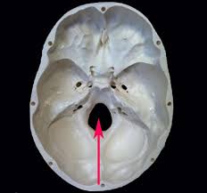
This arrow showing Foramen Spinosum,,Contents are ?
| A |
Vertebral venous plexus |
|
| B |
Vertebral artery |
|
| C |
Middle meningeal artery |
|
| D |
Maxillary nerve |
The lateral segments of the middle fossa are deeper than its middle portion; they support the temporal lobes of the brain and show depressions that mark the convolutions of the brain. These segments are traversed by furrows for the anterior and posterior branches of the middle meningeal vessels, which pass through the foramen spinosum.
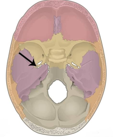
The canal marked by an arrow, is a part of?
| A |
Lesser wing of sphenoid |
|
| B |
Greater wing of sphenoid |
|
| C |
Ethmoid |
|
| D |
Pterygoid |
Ans. is ”A i.e., Lesser wing of sphenoid
The optic nerve leaves the orbit is the optic canal to enter the cranial vault.
The optic canal is the most posterior landmark of the orbit. It measures 10 mm in length.
The thin piece of bone separating the optic canal from the superior orbital fissure is the optic strut.
The optic strut and optic canal are a part of the lesser wing of sphenoid bone
| A | Glossopharyngeal nerve | |
| B |
vagus nerve |
|
| C |
Accessory nerve |
|
| D |
All the above |
The glossopharyngeal nerve, the vagus nerve and the accessory nerve, so cranial nerves IX, X and XI pass through the jugular foramen. That’s four structures to remember. You’ve got cranial nerves IX, X and XI and you’ve got the internal jugularvein.
Structure passing through both greater and lesser sciatic foramen is ‑
| A |
Pudendal nerve |
|
| B |
Sciatic nerve |
|
| C |
Superior gluteal nerve |
|
| D |
Inferior gluteal nerve |
Ans. is ‘a’ i.e., Pudendal nerve
Three structures pass through both greater and lesser sciatic foramen :-
- Pudendal nerve
- Internal pudendal vessels
- Nerve to obturator internus
Structure passing throught greater sciatic foramen
- Piriformis fills the foramen almost completely
- Structures passing above the piriformis
- Superior gluteal nerve
- Superior gluteal vessels
- Structures passing below the piriformis
- Inferior gluteal nerve
- Inferior gluteal vessel.
- Sciatic nerve
- Posterior cutaneous nerve of thigh
- Nerve to quadratus femoris
- Pudendal nerve
- Internal pudendal vessels
- Nerve to obturator internus
Note : Last three structures also enter the lesser sciatic foramen.
- Structures passing through lesser sciatic foramen
- Pudendal nerve
- Internal pudendal vessels
- Nerve to obturator internus
| A | Emissary vein | |
| B |
Vagus nerve |
|
| C |
Mandibular nerve |
|
| D |
Internal jugular vein |
Ans. is ‘c’ i.e., Mandibular nerve
Foramen ovale transmits all except
| A |
Emissary vein |
|
| B |
Mandibular nerve |
|
| C |
Lesser petrosal nerve |
|
| D |
Middle meningeal artery |
Ans. is ‘d’ i.e., Middle meningeal artery
Foramen ovate transmits (mnemonic – MALE) :-
- Mandibular nerve
- Accessory meningeal artery
- Lesser petrosal nerve
- Emissory vein