|
Correct Answer � C
Explanation
|
|
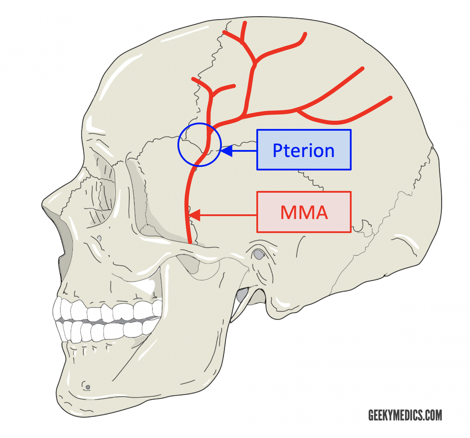
The highlighted structure in the image is the pterion, which is the meeting point of four skull bones: the frontal, parietal, temporal, and sphenoid bones.
This area is clinically significant due to its thinness and proximity to important blood vessels.
The middle meningeal artery, a branch of the maxillary artery, courses beneath this point, but not the accessory middle meningeal artery.
This H-shaped suture is crucial because it harbors the middle meningeal artery, a significant blood vessel that supplies the meninges and the calvaria.
The pterion corresponds to the location of the anterolateral (sphenoidal) fontanelle in the fetal skull, which closes during early childhood.
Important points about the pterion:
Location: The pterion is typically found on the lateral aspect of the skull, approximately 2.5 cm (1 inch) above the zygomatic arch and slightly anterior to the external auditory meatus.
Clinical Importance:
Middle Meningeal Artery: As you mentioned, the middle meningeal artery courses beneath the pterion. It arises from the maxillary artery and enters the cranium through the foramen spinosum. It then runs along the inner surface of the skull, often grooving the adjacent bone, until it reaches the pterion. At this point, it splits into anterior and posterior branches.
Pterional Approach: Neurosurgeons use the pterion as a surgical landmark. The pterional approach involves making an incision over the pterion to access the anterior cranial fossa, the sylvian fissure, and various pathologies (such as aneurysms or tumors).
Accessory Middle Meningeal Artery:
- While the middle meningeal artery is the primary vessel coursing beneath the pterion, there is also an accessory middle meningeal artery in some individuals.
- This accessory artery may arise independently from the maxillary artery or from a separate small foramen near the foramen ovale.
- Unlike the main middle meningeal artery, the accessory version does not necessarily follow the pterion’s course.