

Question
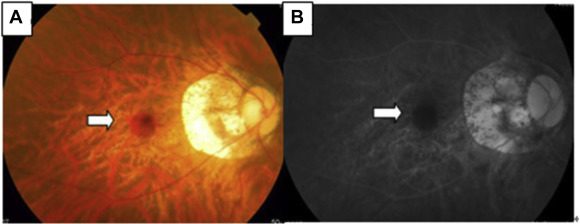
Identify the condition shown in the image?

| A. |
Diabetic retinopathy |
| B. |
Pathological myopia |
| C. |
Sarcoidosis |
| D. |
Hypertensive retinopathy |
Show Answer


|
Correct Answer � B Explanation |
 |

-
- The image shows degenerative changes in Pathological/degenerative myopia.
- Pathological/Degenerative/Progressive myopia usually leads to degenerative changes in the retina along with a high degree of refractive error.
-
Degenerative myopia:
- Refractive error may increase up to 20-25D with degenerative changes in the eye.
- Temporal myopic crescent is a feature of pathological/degenerative myopia.
- It is a white crescent at the temporal border of the disc.
Some of the most typical features of degenerative myopia are:- Vitreous liquefaction and posterior vitreous detachment
- Peripapillary atrophy appearing as temporal choroidal or scleral crescents or rings around the optic disc
- Lattice degeneration in the peripheral retina
- Tilting or malinsertion of the optic disc, usually associated with myopic conus
- Thinning of the retinal pigment epithelium with resulting atrophic appearance of the fundus
- Ectasia of the sclera posteriorly (posterior staphyloma)
- Breaks in Bruch’s membrane and choriocapillaris, resulting in lines across the fundus called “lacquer cracks”
- Foster Fuch’s spot in the macular area.

