

Question
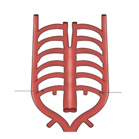
The image shows the derivatives of the embryological aortic arches (pharyngeal arch arteries). Which of the following structures develops from the 6th pharyngeal arch artery on the left side?

| A. |
Arch of Aorta |
| B. |
Ductus arteriosus |
| C. |
Carotid arteries |
| D. |
Subclavian artery |


|
Correct Answer » B Explanation |
 |

The image illustrates the aortic arch system in embryological development, showing the paired pharyngeal (aortic) arch arteries that connect:
-
Ventral aorta (aortic sac)
-
Dorsal aorta
There are six pairs of embryonic aortic arches, although some regress during development.
The 6th aortic arch is also called the pulmonary arch.
-
Right side → proximal right pulmonary artery
-
Left side → proximal left pulmonary artery + ductus arteriosus
This ductus arteriosus connects the pulmonary artery to the descending aorta in fetal circulation.
During embryonic development there are 6 paired aortic arches, though the 5th arch regresses.
| Aortic Arch | Adult Derivatives |
|---|---|
| 1st arch | Maxillary artery |
| 2nd arch | Stapedial and hyoid arteries |
| 3rd arch | Common carotid + proximal internal carotid |
| 4th arch | Right → proximal right subclavian artery |
| Left → arch of aorta | |
| 5th arch | Disappears |
| 6th arch | Right → right pulmonary artery |
| Left → ductus arteriosus |