Question
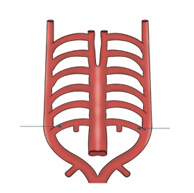
The image shows the derivatives of the embryological aortic arches (pharyngeal arch arteries). Which of the following structures develops from the 6th pharyngeal arch artery on the left side?
| A. |
Arch of Aorta
|
| B. |
Ductus arteriosus
|
| C. |
Carotid arteries
|
| D. |
Subclavian artery
|
Show Answer
|
Correct Answer » B
Explanation
|
|
The image illustrates the aortic arch system in embryological development, showing the paired pharyngeal (aortic) arch arteries that connect:
There are six pairs of embryonic aortic arches, although some regress during development.
The 6th aortic arch is also called the pulmonary arch.
This ductus arteriosus connects the pulmonary artery to the descending aorta in fetal circulation.
During embryonic development there are 6 paired aortic arches, though the 5th arch regresses.
| Aortic Arch |
Adult Derivatives |
| 1st arch |
Maxillary artery |
| 2nd arch |
Stapedial and hyoid arteries |
| 3rd arch |
Common carotid + proximal internal carotid |
| 4th arch |
Right → proximal right subclavian artery |
| |
Left → arch of aorta |
| 5th arch |
Disappears |
| 6th arch |
Right → right pulmonary artery |
| |
Left → ductus arteriosus |