

Question
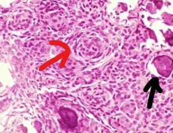
A 48-year-old female presents with a several-month history of persistent headaches. Her husband notes that over the past few months, the patient has seemed apathetic and depressed. On physical exam, the patient’s left leg strength is 4/5, and her gait is unsteady. On MRI, a 5 cm. mass in the parasagittal region of the right frontal lobe is present. Surgery is performed, revealing a multilobular tumor that is easily separated from the underlying brain. A biopsy of the tumor is shown here in the photomicrograph below.What is the diagnosis?

A. Metastatic carcinoma.
B. Meningioma.
C. Glioblastoma.
D. Oligodendroglioma.


|
Correct Answer » B Explanation |
 |

The photomicrograph shown above resembles meningioma.Meningiomas are predominantly benign tumors that arise from the meninges. If you want to be more specific, the cell of origin is the meningothelial cell of the arachnoid. They usually present as rounded, bosselated masses that compress (rather than invade) the underlying brain.
Meningiomas commonly show a syncytial (or “meningothelial”) pattern of growth, which means that the cells sit in tight, whorled clusters. Another clue to the diagnosis in this case is the presence of psammoma bodies (represented by black arrow), which are common in meningiomas. And whorls are represented by a red arrow.Clinically, meningiomas are usually slow-growing, low-grade lesions. For tumors in favorable locations, up to 85% are curable with surgery.