


Question
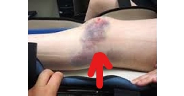
The injury shown in the picture below is best identified using ?

A. Conventional X-ray.
B. CT scan.
C. MRI scan.
D. PET scan.
Show Answer


|
Correct Answer » C Explanation |
 |

Ans:C. MRI Scan
The injury shown in the picture above represents a Bone bruise, or contusion
Bone bruise
It is a traumatic injury to a bone that is less severe than a bone fracture.
- Bones close to the surface of your skin are more likely to be bruised than other bones.
- Bone bruises commonly occur in the knee or femur, but can also occur in the wrist, elbow, heel, foot, hip or pelvis.
- Black and blue mark on skin happens when an injury damages small blood vessels and causes blood and fluid to leak into the nearby tissues and blood vessels.
There are three types of bone bruises:
- Subperiosteal hematoma:With a bone fracture, all of the trabeculae in a region of bone have broken. But with a bone bruise, an injury only damages some of these trabeculae. An injury might cause blood to build up in the area beneath the periosteum. This causes a subperiosteal hematoma
- Subchondral bruise:An injury might also cause bleeding and swelling in the area between your cartilage and the bone beneath it. This causes a subchondral bone bruise.
- Interosseous bruise: bleeding and swelling can happen in the medulla of your bone. This is called an interosseous bone bruise.
Symptoms:
- Stiffness
- Significant pain and swelling
- The pain is more severe than a soft tissue bruise
- Tenderness and pain lasting longer than a usual bruise
- Trouble using an injured joint
Diagnosis:
- A bone bruise or contusion is best identified using MRI scan.
- A bone bruise doesn’t show up on an X-ray.