


Question
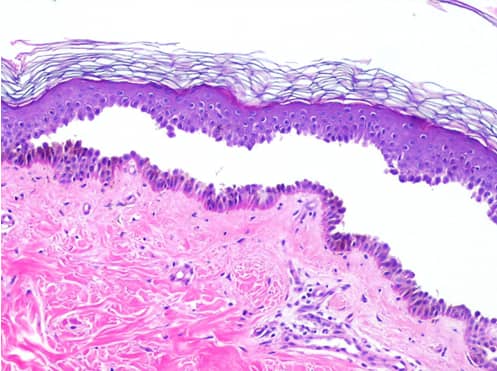
A histopathological image of the skin biopsy of a patient given below is suggestive of:

A. Leprosy
B. Lichen planus
C. Pemphigus vulgaris
D. Psoriasis
Show Answer


|
Correct Answer » C Explanation |
 |

Answer- C. Pemphigus vulgaris
- Histopathological image shown above is showing a suprabasal split between the keratinocytes and basal layer. The attached basal keratinocytes give an appearance of ‘Tomb stoning’. These findings are characteristically seen in Pemphigus vulgaris. Acantholytic cells can be seen at higher power in the blister cavity and the edge of blister.