
Question
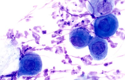
This photomicrograph of this cells shows the presence of?
A. Melanocyte
B. Keratinocyte
C. Neutrophils
D. Monocyte
Show Answer

|
Correct Answer » B Explanation |
 |
The above photomicrograph shows acantholytic cells with the presence of keratinocytes.
The bulla in pemphigus shows presence of acantholytic cells
These cells (which are present both in the blister cavity and at the edge of the blister) are rounded keratinocytes with the cytoplasm condensed in the periphery, resulting in a perinuclear halo
Pemphigus:
- Autoimmune disease
- Antibodies against intercellular substance
- Intradermal acantholytic bullae present (with oral mucosal lesion)
- Mucus membrane lesions seen
- Acantholysis is seen in epidermis
Haley-Haley disease
- It is also known as familial benign pemphigus
- NO sub-epidermal blistering
- Intra-epidermal vesicle caused by breaking apart of epidermal cells
- Definitive diagnosis also requires the demonstration of anti-desmoglein autoantibodies by direct immunofluorescence on the skin biopsy
