ANKLE JOINT- LIGAMENTS
ANKLE JOINT
- Synovial unaxial hinge joint.
- Ankle joint is formed by 3 bones:
– Tibia
– Fibula
– Talus
- The factors contributing to the stability of the ankle joint are:
1. Close interlocking of he articular surfaces
- The shape of the articular surfaces maintain the stability of the joint.
2. Fibrous capsule
- The joint is covered by a thin fibrous capsule which is strengthened medially & laterally by the collateral ligaments.
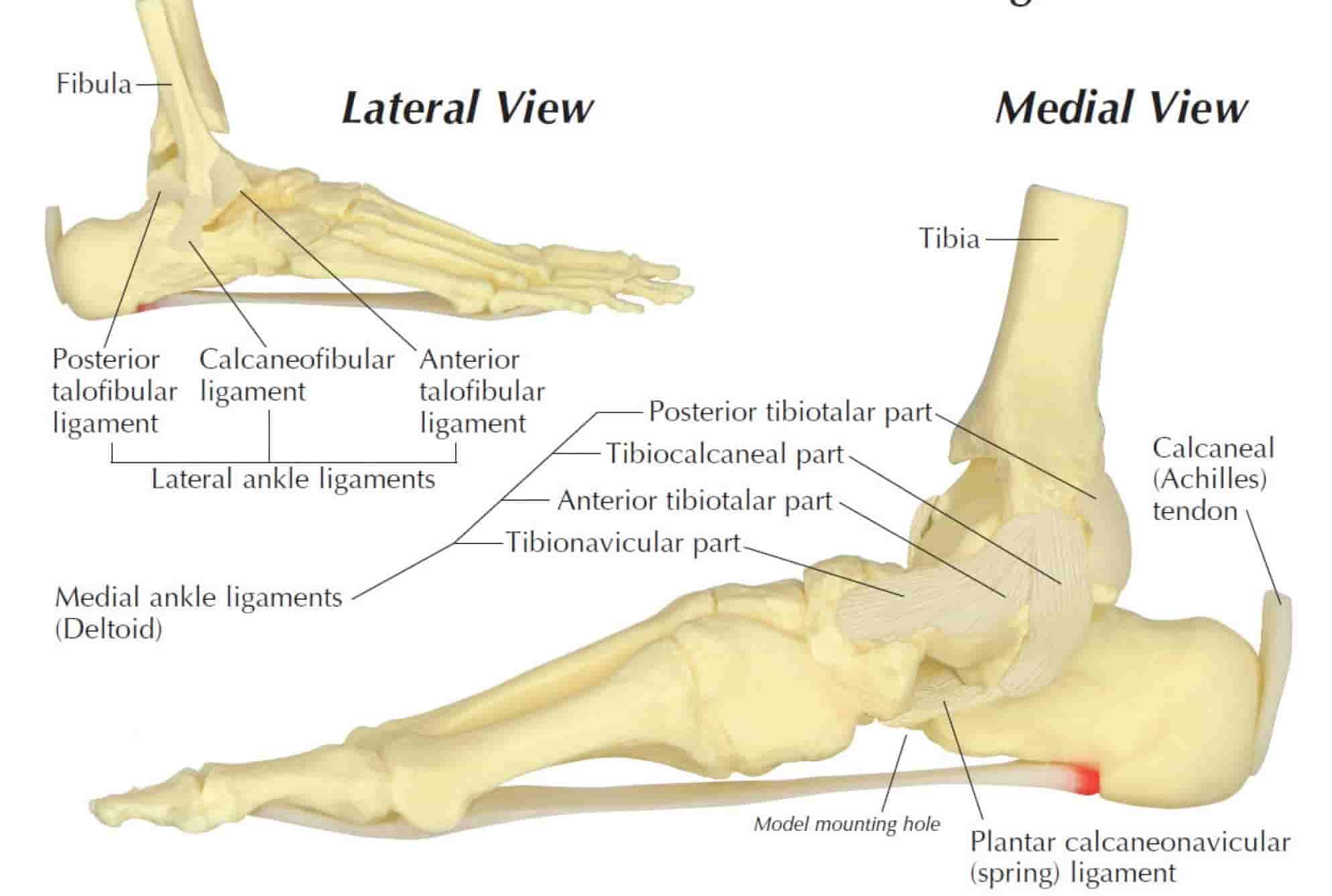
3. Medial collateral ligament (Deltoid ligament)
- Supports the medial side of joint
- Proximally , it is attached to the medial malleolus.
Distally, it is attached to four places:
- Sustentaculum tali of calcaneus
- Calcaneonavicular ligament(support head of talus)
- Navicular tuberosity
- Medial surface of talus
– Deltoid ligament is a strong, flat, triangular band, attached, above to the apex and anterior and posterior borders of medial malleolus.
Deltoid ligament is composed of :
1. Anterior Tibiotalar Ligament
2. Tibiocalcaneal Ligament: Attached to sustenticulum tali of calcaneum.
3. Posterior Tibiotalar Ligament: Attached to medial tubercle & medial surface of talus.
4. Tibionavicular Ligament: Attached to navicular tuberosity & spring ligament.
- It consists of two sets of fibers, superficial and deep.
- Its middle portion, together with the calcaneofibular ligament, binds the bones of the leg firmly to the foot, and resists displacement in every direction.
- Its anterior and posterior fibers limit extension and flexion of the foot respectively, and the anterior fibers also limit abduction
- Ligamentous support is more important during plantar flexion.
- Ankle joint is more stable at dorsi-flexed position
- Deltoid ligament , Lateral ligament, Shape of the superior talar articular surface stablizes ankle joint
- It usually resists a force which fractures the malleolus, to which it is attached.
4. Lateral collateral ligaments
- Anterior and posterior talofibular ligaments support the lateral side of the joint from lateral malleolus to the posterior and anterior ends of talus.
- Anterior talofibular ligament (most commonly affected on ankle joint injury).
- Though it does not span across the ankle joint itself, the syndesmotic ligament makes an important contribution to the stability of ankle.
- This ligament spans the syndesmosis (the articulation between the medial aspect of distal fibula and the lateral aspect of the distal tibia.
- An isolated injury to this ligament is often called high ankle sprain.
- Calcaneofibular ligament is attached at the lateral malleolus and to the lateral surface of calcaneum.
Exam Important
- Deltoid ligament is attached to medial malleolus, Sustentaculum tali of calcaneus, Calcaneonavicular ligament, Navicular tuberosity, Medial surface of talus
- Deltoid ligament strengthens ankle joints
- Ankle joint is more stable at dorsi-flexed position
- Deltoid ligament , Lateral ligament, Shape of the superior talar articular surface stablizes ankle joint
- Anterior talofibular ligament gets affected most commonly in injury of ankle
- Plantar calcaneonavicular ligament support head of talus.
Don’t Forget to Solve all the previous Year Question asked on ANKLE JOINT- LIGAMENTS
Click Here to Start Quiz
Click Here to Start Quiz
