ASTIGMATISM
Assertion:
The slit-lamp examination of the eye will show thinning and ectasia of the central cornea.Reason: Slit-lamp examination findings are consistent with keratoconus.
| A | Both Assertion and Reason are true, and Reason is the correct explanation for Assertion | |
| B |
Both Assertion and Reason are true, and Reason is not the correct explanation for Assertion |
|
| C |
Assertion is true, but Reason is false |
|
| D |
Assertion is false, but Reason is true |
A patient presents to the clinic with progressive myopia and irregular astigmatism which does not improve fully with glasses. O/e window reflex is disoriented and placcid disc examination shows irregularities of the cornea.
Assertion: The slit-lamp examination of the eye will show thinning and ectasia of the central cornea.
Reason: Slit-lamp examination findings are consistent with keratoconus.
| A |
Both Assertion and Reason are true, and Reason is the correct explanation for Assertion |
|
| B |
Both Assertion and Reason are true, and Reason is not the correct explanation for Assertion |
|
| C |
Assertion is true, but Reason is false |
|
| D |
Assertion is false, but Reason is true |
Keratoconus is a noninflammatory bilateral ecstatic condition of the cornea in its axial part. Slit-lamp examination in this condition shows thinning and ectasia of the central cornea, presence of opacity at the apex, Fleischer’s ring at the base of the cone, folds in the Descemet’s membrane and Bowman’s membrane.
In simple astigmatism, foci of image formed on:
| A |
Both image are formed in front of retina |
|
| B |
Both image are formed behind retina |
|
| C |
One image front & other behind the retina |
|
| D |
One on retina, other behind the retina |
D. i.e. One on retina, other behind the retina
In simple astigmatism, one image is formed upon retina (i.e. one meridian is emmetropic) and the other image may be formed behind or in front of the retinaQ (i.e. other meridian is either hypermetropic or myopic). These are designated as simple hypermetropic and simple myopic astigmatism respectively.
In compound astigmatism, both images are formed either in front (k/a compound myopic astigmatism) or behind retina (k/a compound hypermetropic astigmatism). And in mixed astigmatism one image is formed in front and other behind the retina.
Astigmatism
It is a type of refractory error wherein the refraction varies in the different meridian; so the rays of light entering in the eye cannot converge to a point focus (but form focal lines) upon retina. It can be classified in various ways.
| A |
Vertical meridian is more curved than the horizontal |
|
| B |
Horizontal meridian is more curved than the vertical |
|
| C |
Both meridia are equally curved |
|
| D |
None of the above |
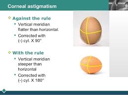
Against-the-rule astigmatism (ATR): The horizontal meridian is more curved than the vertical meridian.
Astigmatism:
- Refractive error in which the refraction varies in different meridians of the eye, due to which light rays fail to converge in a point focus.
- In this condition, there is a difference in refractive powers in the two principal meridians of the eye.
- As a result, the rays in the two meridians are focused at different points leading to a blurred image. Based on the axis of the two principal meridians, astigmatism is of two main types:
Regular Astigmatism:
- Here the two meridians are perpendicular to each other.
- If the horizontal axis is steeper than vertical, it is called “against the rule” astigmatism.
- If the vertical is steeper than horizontal, it is called “with the rule” astigmatism.
- If the axes are perpendicular but obliquely inclined, it is called oblique astigmatism.
-
Bi-oblique astigmatism: Here, the two principal meridians are not at right angles to each other; for example, one
may be at 40 degrees and the other, at 100 degrees.
Astigmatism is considered to be:
| A |
Spherical abberation |
|
| B |
Curvatural ametropia |
|
| C |
Axial ametropia |
|
| D | Index ametropia |
Ans. Spherical abberation
Lens used to treat astigmatism:
September 2005
| A |
Concave lens |
|
| B |
Spherical lens |
|
| C |
Convex lens |
|
| D |
Cylindrical lens |
Ans. D: Cylindrical lens
Corneal astigmatism is created when the cornea is not a perfect sphere. The astigmatic cornea is curved more in one meridian than it is in the other. These meridians are usually 90 degrees apart (regular astigmatism).
The lens can have astigmatism also, this is termed lenticular astigmatism. Lenticular astigmatism is evident when there is a significant difference between the astigmatism as measured on the keratometer and refractive astigmatism Astigmatism is corrected optically with a cylindrical lens.
A combination of a spherical lens and a cylindrical lens (spherocylindrical lens) is used to correct a spherical error with an astigmatic error.
| A | Perpendicular principal meridians | |
| B |
Non perpendicular principal meridians |
|
| C |
Any of the above |
|
| D |
None of the above |
Ans. is ‘b’ i.e., Non perpendicular principal meridians
ASTIGMATISM
- Astigmatism is a type of refractive error wherein the refraction varies in the different meridia. Consequently, the rays of light entering in the eye cannot converge to a point focus but form focal lines. The refractive error of the astigmatic eye stems from a difference in degree of curvature refraction of the two different meridians (i.e., the eye has different focal point in different planes). For example, the image may be clearly focused on retina in the horizontal plane, but not in the vertical plane.
- The most common cause of astigmatism is abnormality of corneal curvature. Other less common causes are lenticular (curvature abnormality of lens,oblique position of lens) and retinal (oblique placement of macula).
Types of astigmatism
A) Based on axis of the principal meridians
1) Regular stigmatism : Principal meridians are perpendicular
- With-the-rule astigmatism-the vertical meridian is steepest.
- Against-the-rule astigmatism-the horizontal meridian is steepest.
- Oblique astigmatism-the steepest curve lies in between 120 and 150 degrees and 30 and 60 degrees.
2) Irregular astigmatism – principal meridians are not perpendicular.
B) Based on focus of the principal meridians
1) Simple astigmatism
- Simple hyperopic astigmatism – first focal line is on retina while the second is located behind the retina
- Simple myopic astigmatism – first focal line is in front of the retina while the second is on the retina.
2) Compound astigmatism
- Compound hyperopic astigmatism-both focal lines are located behind the retina.
- Compound myopic astigmatism-both focal lines are located in front of the retina.
3) Mixed astigmatism-focal lines are on both sides of the retina (straddling the retina).
Treatment ofAstigmatism
- Treatment of astigmatism consists : –
- Optical treatment : – It consists of cylindrical power spectacles or contact lens. Types of contact lenses used are permeable contact lens, Soft toric contact lens (for high degree astigmatism), hybrid lens, i.e., soft on hard lens, hard contact lens.
- Surgical treatment : – Non-laser (astigmatic keratotomy) or laser (PRK, LASIK).
| A |
Astigmatism in which the principal meridians are parallel |
|
| B |
Astigmatism in which the principal meridians are perpendicular |
|
| C |
Asymptomatic astigmatism |
|
| D |
Astigmatism as a result of cataract surgery |
- Regular stigmatism → Principal meridians are pendicular
- Irregular astigmatism → Principal meridians are not perpendicular.