
CARCINOMA OF URINARY BLADDER
CARCINOMA OF URINARY BLADDER
- More common in whites, higher social- economic status.
- 75% tumours are localised to bladder
- 25% spread to regional nodes

CLASSIFICATION-
I) According to histologically-
a) Transitional cell carcinoma (90%)
b) Squamous cell carcinoma
c) Adenocarcinoma
d) Mixed
e) Undifferentiated
ETIOLOGY-
- Transitional cell carcinoma
- Cigratte smoking (MC)
- Chemical carcinogens- naphthylamine benzidine, aniline, acrolein, hydrocarbons (BHAAM)
- Schistosomiasis
- Drugs- phenacetin, chlornaphazine, cyclophosphamide, analgesic
- Pelvic irradiation
- Occupation- rubber, leather etc
2. Squamous cell carcinoma
- Schistosomiasis
- Chronic UTI
- Bladder diverticula
3. Adenocarcinoma (5%)
- Urahal remnants
- Ectopia vesicae
- Occurs in intestinal urinary conduits, augmentation, pouches and ureterosigmoidotomies
FEATURES-
- Transitional Cell carcinoma
- MC type of bladder tumour
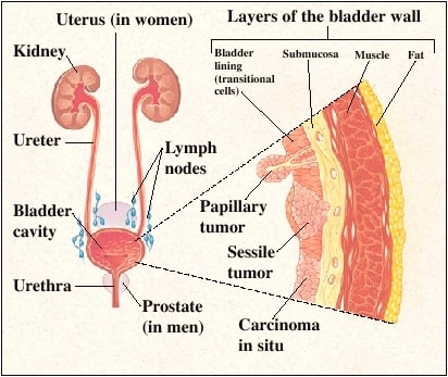
- MC appears as papillary or exophytic lesions
- Carcinoma in situ or flat urotheal carcinoma
- Pagetoid spread
- Sites- lateral wall (MC), trigone
2. Squamous Cell carcinoma-
- Arise from lateral bladder wall and dome
- Nodular and invasive
3. Adenocarcinoma
- 3 types- primary vesical, urchal, metastatic
- Primary adenocarcinoma arise along the floor
- Adenocarcinoma arising from urachus occurs at the dome.
PATHOLOGY-
- MC type grossly in Papillary
- MC type histologically Transitional cell carcinoma
CLINICAL FEATURES-
- Most tumours develop at trigone and adjacent posterolateral wall with urethral involvement
- Tumour tend to be multifocal in bladder
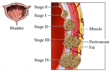
STAGING-

INVESTIGATIONS-
- Cystoscopy (gold standard investigations)-
- Cystoscopy and transurethral resection (TUR) is for diagnosis
- Detecting cancer in symptomatic patient and assessing response to treatment
- Most useful for early diagnosis of recurrence in TCC
2. IVP-
- Filling defect in bladder, dilatation of ureter
3. Ultrasound-
- It is useful investigation which can detect a bladder carcinoma
- Can detect liver metastases
4. CT scan is IOC for staging
5. Bimanual palpation-
- Rectoabdominally in males
- Vaginoabdominally in females
6. New tests-
- Detection of antigens such as nuclear matrix proteins (NMP22) or mini chromosome maintence (MCM) in urine
- Detect new or recurrent tumours

TREATMENT-
- Stage Tis, Ta, T1
- Transurethral resection of tumour (Ta)
- Chemotherapy- thiotepa (MC- BCG)/ adriamycin/ mitomycin
- BCG or interferon immunotherapy (Tis)
- T1- TUR + BCG + chemotherapy
2. T2- T4 lesion
- Radical cystectomy followed by chemotherapy
- Neoadjuvant chemotherapy followed by Radical cystectomy
3. Any T, N1, M0 or any T, N0, M1
- Systemic chemotherapy
4. Small lesions
- Partial cystectomy
Exam Important
- Transitional cell carcinoma
- Cigratte smoking (MC)
- Chemical carcinogens- naphthylamine benzidine, aniline, acrolein, hydrocarbons (BHAAM)
- Schistosomiasis
- Drugs- phenacetin, chlornaphazine, cyclophosphamide, analgesic
- Pelvic irradiation
- Occupation- rubber, leather etc
2. Squamous cell carcinoma
- Schistosomiasis
- Chronic UTI
- Bladder diverticula
3. Adenocarcinoma (5%)
- Urahal remnants
- Ectopia vesicae
- Occurs in intestinal urinary conduits, augmentation, pouches and ureterosigmoidotomies
FEATURES-
- Transitional Cell carcinoma
- MC type of bladder tumour
- MC appears as papillary or exophytic lesions
- Carcinoma in situ or flat urotheal carcinoma
- Pagetoid spread
- Sites- lateral wall (MC), trigone
2. Squamous Cell carcinoma-
- Arise from lateral bladder wall and dome
- Nodular and invasive
3. Adenocarcinoma
- 3 types- primary vesical, urchal, metastatic
- Primary adenocarcinoma arise along the floor
- Adenocarcinoma arising from urachus occurs at the dome.
PATHOLOGY-
- MC type grossly in Papillary
- MC type histologically Transitional cell carcinoma
- Cystoscopy (gold standard investigations)-
- Cystoscopy and transurethral resection (TUR) is for diagnosis
- Detecting cancer in symptomatic patient and assessing response to treatment
- Most useful for early diagnosis of recurrence in TCC
2. IVP-
- Filling defect in bladder, dilatation of ureter
3. Ultrasound-
- It is useful investigation which can detect a bladder carcinoma
- Can detect liver metastases
4. CT scan is IOC for staging
5. Bimanual palpation-
- Rectoabdominally in males
- Vaginoabdominally in females
6. New tests-
- Detection of antigens such as nuclear matrix proteins (NMP22) or mini chromosome maintence (MCM) in urine
- Detect new or recurrent tumours
TREATMENT-
- Stage Tis, Ta, T1
- Transurethral resection of tumour (Ta)
- Chemotherapy- thiotepa (MC- BCG)/ adriamycin/ mitomycin
- BCG or interferon immunotherapy (Tis)
- T1- TUR + BCG + chemotherapy
2. T2- T4 lesion
- Radical cystectomy followed by chemotherapy
- Neoadjuvant chemotherapy followed by Radical cystectomy
3. Any T, N1, M0 or any T, N0, M1
- Systemic chemotherapy
4. Small lesions
- Partial cystectomy
Click Here to Start Quiz

