Dermatitis
Which of the following cause allergic contact dermatitis through air?
| A | House dust mite | |
| B |
Nickel |
|
| C | Parthenium | |
| D |
Detergents |
Which of the following cause allergic contact dermatitis through air?
| A | House dust mite | |
| B |
Nickel |
|
| C | Parthenium | |
| D |
Detergents |
Parthenium
REF: Roxburgh’s 17th ed page 105-109, rook’s- 7th ed p. 19.1-19.20, fitzpatrick’s p. 1181-1203 CONTACT DERMATITIS
- Type 4 hypersensitivity
- Diagnosis by patch test (read @ 2 days)
- Most common air borne contact dermatitis: Parthenium (congress grass)
- Most common contact dermatitis in indian females: Detergent dermatitis
- Most common contact dermatitis due to metal: Nickel
Most common cause of plant induced dermatitis in India:
| A | Poison ivy | |
| B | Parthenium | |
| C |
Ragweed |
|
| D | Cotton fibres |
“Most common cause of plant induced dennatitis is parthenium.”
Parthenium hysterophorus (congress grass, congress weed carrot weed, wild fever few, the “scourge of India”)
- It is an exotic weed that was accidently introduced in India in 1956 through imported food grains.
- It has become a common weed causing dermatitis of epidemic proportions.
- The epithet “congress weed” refers to the U.S. congress (who allocated the shipment for Pune, India).
- In Pune it found an ecological niche without natural enemies and spread rapidly along the canal banks, roads and railway tracks to become a major field weed.
- Both rural and urban areas have been invaded by this weed.
- It is the leading cause of plant induced air borne contact dermatitis in India and has achieved major weed status in India and Australia within the past few decades.
- The weeds can affect human health, animal husbandry, crop production and biodiversity.
A 3 year old child has eczematous dermatitis on extensor surfaces. His mother has a history of Bronchial asthma. Diagnosis should be
| A | >Atopic dermatitis | |
| B | >Contact dermatitis | |
| C | >Seborrhic dermatitis | |
| D | >Infantile eczematous dermatitis |
Atopic dermatitis [Harrison 16th/e p 268, Roxburgh 17th/e p 105-112]
- Eczematous dermatitis on extensor surface and the history of bronchial asthma suggests the diagnosis of Atopic dermatitis.
Atopic dermatitis
Etiology and pathogenesis
- Genetic predisposition is very important but the precise mode of inheritence is uncertain.
- Atopic diseases tend to run true to type within each family. In some families the affected members predominantly have eczema while in others respiratory problems predominate.
This is probably because the dermatitis and asthma are inherited through separate though closely related genetic pathways.
Immunological changes
- Elevated /gE level seen in 80% of the patients.
- Abnormalities in lymphocytes are also seen. Clinical features
Clinical picture varies with the age of the patient
(i) Infantile
- Begins at about 3 months
- Severely itchy exudative lesions on face and other parts.
(ii) Childhood
- Itch leathery flexural lesions, sometimes extensor lesions occur.
(iii) Adult
- Lichnefied itchy lesions
- Seen in cubital and popliteal fossa.
Seborrhic dermatitis
- Caused by an yeast, Malassezia futfur
- Usually seen in adults, sometimes in infants but not in children.
- The lesions involve the scalp, face (nasolabial folds, eyebrows and eyelashes) presternal and interscapular regions and major flexures (axilla, groin and inframammary region) umbilicus and natal cleft. This distribution of seborrhic dermatitis is very characteristic and is called “seborrhic distribution”.
Contact dermatitis
- It develops due to contact with chemicals.
| A | Scalp | |
| B |
Elbow |
|
| C |
Ante-cubital fossa |
|
| D |
Trunk |
Ans:C.)Antecubital Fossa.
Atopic dermatitis
- It is a chronic, pruritic inflammatory skin disease (see image below) of unknown origin that usually starts in early infancy, but also affects a substantial number of adults. AD is commonly associated with elevated levels of immunoglobulin E (IgE). That it is the first disease to present in a series of allergic diseases—including food allergy, asthma, and allergic rhinitis, in order—has given rise to the “atopic march” theory.
- The earliest lesions affect the creases (antecubital and popliteal fossae), with erythema and exudation. Over the following few weeks, lesions usually localize to the cheeks, the forehead and scalp, and the extensors of the lower legs; however, they may occur in any location on the body, usually sparing the diaper area and the nose. Lesions are ill-defined, erythematous, scaly, and crusted (eczematous) patches and plaques.
| A |
Dennie -Morgan fold |
|
| B |
Hertoghe’s sign |
|
| C |
Darier’s Sign |
|
| D |
Hyperlinearity of palms |
It is seen in urticaria pigmentosa.
All are skin findings in atopic dermatitis, EXCEPT:
| A |
Facial pallor |
|
| B |
Pityriasis alba |
|
| C |
Keratosis pilaris |
|
| D |
Black dermatographism |
First 3 options are included under the minor criteria for atopic dermatitis.
In atopic dermatitis white dermatographism occures which is a white line occuring in the skin on stroking with a blunt object.
Black dermaotographism is discolouration of the skin after pressure from a metallic object.
Ref: Fitzpatrick’s Dermatology in General Medicine, 7th edition, Page : 148
Which of the following conditions produce a seborrheic dermatitis like lesions in an infant?
| A |
Langerhan cell histiocytosis |
|
| B |
Juvenile xanthogranuloma |
|
| C |
Multicentric histiocytosis |
|
| D |
Erdheim Chester disease |
All the above mentioned optionscare disorders of histiocytes.
In langerhan cell histiocytosis, bone, lymph node and skin are the most frequently affected sytems.
The most characteristic presentation is with scalp involvement.
which is erythematous with greasy scales, looking very like seborrheic dermatitis.
S100 Paraffin, peanut agglutinin, Placental alkaline phosphatase are the common used immunological markers.
Air-borne contact dermatitis can be diagnosed using which of the following investigation?
| A |
Skin biopsy |
|
| B |
Patch test |
|
| C |
Prick test |
|
| D |
Estimation of serum IgE levels |
36-year-old farmer presented to dermatology department with pruritic erythematous lesions on arms, forearms, face and retro auricular area after removal of weeds in his farm. A diagnosis of phytodermatitis was made. What is the most likely plant responsible for this condition?
| A |
Parthenium hysterophorus |
|
| B |
Urtica diodica |
|
| C |
Alstromecia |
|
| D |
Melaleuca alternifolia |
Parthenium hysterophorus or congress weed is the most common plant producing phytodermatitis in India.
It presents with pruritic skin lesions on exposed areas even involving the photoprotected areas.
Hence called pseudophotodermatitis.
Allergen responsible is a sesquiterpene lactone.
Which of the following is the commonest air borne contact dermatitis in India?
| A |
Parthenium hysterophorus |
|
| B |
Pottasium dichromate |
|
| C |
Nickel suphate |
|
| D |
Toxicodendron radicans |
Parthenium is the most common contact sensitizer (20%), followed by potassium dichromate (16%), xanthium (13.3%), nickel sulfate (12%), chrysanthemum (8%), mercaptobenzothiazole and garlic (6.7% each) in the study with Indian Standard Series and indigenous antigens.
Important Note: Appraisal of Best National Studies
Parthenium dermatitis is an immuno-inflammatory disease caused by Parthenium hysterophorus and is the commonest cause of plant dermatitis in India. It is caused by airborne dry and friable plant particles including trichomes, and the most important allergens responsible for allergic contact dermatitis are sesquiterpene lactones. It is responsible for 40% of patients attending contact dermatitis clinics.
Hypersensitivity: Type IV and Type I combined. Contact sensitivity to parthenium is long term, and hence the disease runs a chronic course with exacerbation during summers.
Corticosteroids are the mainstay of treatment but the prolonged use of corticosteroids can cause serious side effects. Azathioprine used in daily doses has been shown to be effective.
Toxicodendron radicans : Is Poison Ivy, the commonest herbal contact allergen in United States.
Ref: Indian J Dermatol Venereol Leprol. 2012 Sep; 78 (5) :560-8: Parthenium dermatitis in India: past, present and future.
Sharma VK, Verma P, Department of Dermatology and Venereology, AIIMS, New Delhi, India.
Verma KK, Bansal A, Sethuraman G: Parthenium dermatitis treated with azathioprine weekly pulse doses. Indian J Dermatol Venereol Leprol 72:24, 2006
J Dermatol. 2000 Jul;27 (7) :440-5; Common contact sensitizers in Delhi. Singhal V, Reddy BS. Source Department of Dermatology and STD, Maulana Azad Medical College & Loknayak Hospital, New Delhi, India.
| A | Atopic dermatitis | |
| B |
Contact dermatitis |
|
| C |
Seborrheic dermatitis |
|
| D |
Infantile eczematous dermatitis |
Diagnostic criteria for atopic dermatitis must include pruritus, typical morphology and distribution (flexural lichenification, hand eczema, nipple eczema, and eyelid eczema in adults), onset in childhood, and chronicity.
| A |
Leukemia |
|
| B |
Lymphoma |
|
| C |
Histiocytosis X |
|
| D |
Multiple myeloma |
LCH provokes a non-specific inflammatory response, which includes fever, lethargy, and weight loss. Organ involvement can also cause more specific symptoms.
Bone: The most-frequently seen symptom in both unifocal and multifocal disease is painful bone swelling. The skull is most frequently affected, followed by the long bones of the upper extremities and flat bones. Infiltration in hands and feet is unusual. Osteolytic lesions can lead to pathological fractures.
Skin: Commonly seen are a rash which varies from scaly erythematous lesions to red papules pronounced in intertriginous areas. Up to 80% of LCH patients have extensive eruptions on the scalp.
Bone marrow: Pancytopenia with superadded infection usually implies a poor prognosis. Anemia can be due to a number of factors and does not necessarily imply bone marrow infiltration.
Lymph node: Enlargement of the liver in 20%, spleen in 30% and lymph nodes in 50% of histiocytosis cases.
Endocrine glands: Hypothalamic pituitary axis commonly involved. Diabetes insipidus is most common. Anterior pituitary hormone deficiency is usually permanent.
Lungs: Some patients are asymptomatic, diagnosed incidentally because of lung nodules on radiographs; others suffer from chronic cough and shortness of breath.
| A |
Type-I hypersensitivity |
|
| B |
Type-I1 hypersensitivity |
|
| C |
Type-III hypersensitivity |
|
| D |
Type-IV hypersensitivity |
Ans. is ‘d’ i.e., Type IV hypersensitivity
In contact dermatitis which cells play major role
| A |
T- cells |
|
| B |
B-cells |
|
| C |
Langhans cells |
|
| D |
Macrophage |
Ans. is ‘a’ i.e., T- cells
Contact dermatitis is type IV (delayed) hypersensitivity reaction which is mediated by T cells.
The following has maximum propensity for photodermatitis-
| A |
Oxytetracycline |
|
| B |
Doxycycline |
|
| C |
Minocycline |
|
| D |
All |
Ans. is ‘b’ i.e., Doxycycline
o Photodermatitis is caused by demeclocycline (max) and doxycycline.
Minor clinical feature in diagnosis of atopic dermatitis A/E
| A |
Dry skin |
|
| B |
Pruritus |
|
| C |
Morgagnian fold |
|
| D |
Pitriasis alba |
B i.e. Pruritis
Atopic Dermatitis is diagnosed by:
| A |
Patch test |
|
| B |
Wood Lamp |
|
| C |
Clinical Examination |
|
| D |
-IgE |
C i.e. Clinical Examination
Itch (or pruritis)Q is a major diagnostic criteria whereas, Denny Morgan infra orbital fold is a minor criteria for diagnosis of atopic dermatitis.
Atopic dermatitis most commonly involves flexural surfaces like antecubital and popliteal fossa Q. However, in infantile phase, face and extensor surface (convexities) are more commonly involved. Spongiosis of epidermis Q is the histological hallmark of dermatitis (eczema).
Contact dermatitis is diagnosed by patch testQ & Atopic dermatitis is diagnosed by clinical examinationQ. Clinical criteria for diagnosis of atopic dermatitis
- Family h/o allergy/atophy
- Personal history of allergy/atophy i.e. presence of other atopic condition asQ – rhinitis, hay fever, asthma, food allergy or eczemaQ.
- Extremily pruritic lesions commonly on antecubital or popliteal fossaf2
- Dienny morgan foldQ, white dermographism
- Exacerbation & remissions.
- Pruritis and scratching made worse by environmental alteration, changes in temperature, sundry (in rainy season) & rough (woolen) clothingQ and leading to excoriation, lichenificationQ, dryness & Dennie’s line.
- Clinical course lasting longer than 6 weeksQ.
- Lesions typical of eczematous dermatitis i.e. papules, erythematous macules and vesicles, which can coalesce to form patches and plaques.
|
Disease |
Diagnosis made by |
|
Atopic Dermatitis |
Clinical ExaminationQ |
|
Contact Dermatitis |
Patch TestQ |
|
Donovanosis |
MicroscopyQ (demonstration of Donovan bodies or safety pin appearance)Q |
|
Syphilis |
Dark field microscopyQ, FTA-ABS, VDRL, MHA-TP, TPI |
|
Chancroid |
Gram stainine (gram -ve cocco‑ bacilli, school of fish or rail road appearance)Q |
|
LGV |
– Microscopic examination of giemsa stained scrapings for inclusion or elementary bodies – Culture, ELISA |
|
Tinea (Dermatophytes) |
KOH SmearQ |
|
Lupus vulgaris |
BiopsyQ |
| A | Gold | |
| B |
Nickle |
|
| C |
Silver |
|
| D |
Iron |
B i.e. Nickle
Most common cause of allergic contact dermatitis in Indian female is
| A |
Vegetables |
|
| B |
Nail polish |
|
| C |
Detergents |
|
| D |
Dyes |
C i.e. Detergents
Berloque dermatitis is due to contact with –
| A |
Metals |
|
| B |
Cosmetics |
|
| C |
Food |
|
| D |
Plants |
B i.e. Cosmetics
Berlock dermatitis is a phototoxic reaction due to bergamot fragrance oil (5 methoxy psoralen) present in some perfumes & cosmeticsQ. It causes erythema followed by prolonged hyperpigmentation after exposure to sun light.
A 55-year-old male, with uncontrolled diabetes mellitus and hypertension, developed severe air-borne contact dermatitis. The most appropriate drug for his treatment would be:
| A |
Systemic corticosteroids |
|
| B |
Thalidomide |
|
| C |
Azathioprine |
|
| D |
Cyclosporine |
C i.e. Azathioprine
In contact dermatitis, the allergic reaction is mediated by delayed hypersensitivity (cell mediated immunity). It is treated by:
– Antihistamincis
– Steroids (topical/systemic)
– Agents depressing cell mediated immunity as cyclosporine, tacrolimus, azathioprine
| A |
Skin biopsy |
|
| B |
Patch test |
|
| C |
Prick test |
|
| D |
Estimation of serum IgE levels |
B i.e. Patch Test
Exfoliative dermatitis is seen in all the following except
| A |
Pityriasis rosea |
|
| B |
Pityriasis rubra pilaris |
|
| C |
Psoriasis |
|
| D |
Drug hypersensitivity |
A i.e. Pityriasis rosea
Gold poisioning leading to exfoliative dermatitis is treated by :
| A |
Chloroquin |
|
| B |
Steroid |
|
| C |
Antibiotics |
|
| D |
Antihistaminics |
B i.e. Steroid
Exfoliative dermatitis or erythroderma is caused by dermatitis (eczema), psoriasis, lichen planus, pityriasis rubra pilaris and drugs like gold but not by pityriasis rosea and versicolor. It is treated by steroidsQ.
Photosensitive dermatitis is/are :
| A |
Psoriasis |
|
| B |
Pellagra |
|
| C |
Pemphigus |
|
| D |
All |
All i.e. Psoriasis, Pellagra, Pemphigus, SLE, Congenital erythropoietic porphyria
Due to-
Psoriasis,
Pellagra,
Pemphigus,
SLE,
Congenital erythropoietic porphyria
M.C. site of Atopic dermatitis
| A |
Scalp |
|
| B |
Elbow |
|
| C |
Antecubital fossa |
|
| D |
Trunk |
C i.e. Antecubital fossae
Atopic dermatitis may be associated with
| A |
Conjunctivitis |
|
| B |
Keratoconus
|
|
| C |
Cataract |
|
| D |
All of the above |
Ans. All of the above
- Atopic dermatitis (AD) is a widespread, chronic and progressive skin disorder characterised by erythema with oedema, vesicles, and oozing lesions in the acute stage and skin thickening (lichenification) in the chronic stage.
- Ocular complications are more prevalent in individuals with AD compared to the general population and can cause notable morbidity.
- Ocular complications associated with AD, including blepharitis, keratoconjunctivitis, keratoconus, glaucoma, cataracts, retinal detachment, ophthalmic herpes simplex virus infections, and dupilumab-associated ocular complications.
- It is important for dermatologists to be aware of the signs and symptoms of these ocular complications, as timely diagnosis and treatment can prevent irreversible vision loss.
| A |
Dermatitis herpetiformis |
|
| B |
Atopic dermatitis |
|
| C |
Leprsoy |
|
| D |
TB |
Ans. B i.e. Atopic dermatitis
Atopic dermatitis
- Also known as Itch disease
- Childhood and adolescent pattern of atopic dermatitis is marked by:
– Flexural skin dermatitis
– Particularly in the ante-cubital fossa/ popliteal fossa
- Other features of AD:
– Perioral pallor,
– Dennies line,
– Increased palmar markings etc.
- The best test to diagnose AD: Clinical examination
- Berloque dermatitis is due to Cosmetics
Which of the following best describes this inflammatory response?
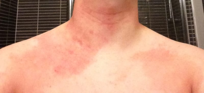
| A | Erythema nodosum | |
| B |
Pemphigus |
|
| C |
Psoriasis |
|
| D |
Spongiotic dermatitis |
RAST test is used in diagnosis of
| A |
Allergic dermatitis |
|
| B |
Seborrhoeicdematitis |
|
| C |
Mycosis fungoides |
|
| D |
Squamous cell carcinoma |
Ans. is ‘a’ i.e., Allergic dermatitis
RAST : Radioallergosorbent assay
- It is the method used to measure total as well as specific IgE against a particular allergen or a complex.
Diagnostic tests in allergic contact dermatitis
- Diagnostic Tests (if indicated)
- Patch testing
- Photopatch testing
- Tests for immediate hypersensitivity
Radioallergosorbent assay test (RAST)
- Open and semiopen patch tests (read at 10 and 45 minutes)
- Prick test
- Scratch-chamber test
- Repeat open application “use” test
- Potassium hydroxide examination to fungi, glass fibers
- Fungal, bacterial, and viral smears and cultures
- Skin biopsies
- Dimethylglyoxime test for detecting nickel, other tests (detection of chromates and formaldehyde)
- Chemical analysis
| A | Atopic dermatitis | |
| B |
Contact dermatitis |
|
| C |
Urticaria |
|
| D |
Erythroderma |
Ans. is ‘a’ i.e., Atopic dermatitis
- Hanifin and Rajka criteria is for diagnosis of atopic dermatitis.
Diagnostic criteria (Hanifin and Rajka)
Based mainly on clinical experience
Major criteria
- Family history of atopy
- Chronicity
- Pruritus
- Typical morphology and distribution
Minor criteria
- Dry skin
- Chelitis
- Elevated edge
- Dennie’s line/dennie morgan fold (infra orbital fold)
- White dermographism
- Peripheral eosinophillia
- Immediate (type i) hypersensivity
- Facial pallor, orbital darkening
- Food intolerance
- Conjunctivitis (recurrent), keratoconus, cataract
- Pityriasis alba
- Hand dermatitis
- Recurrent infections
- At least 3 major or 2 major plus 2 minor criteria are necessary for diagnosis