
EWING SARCOMA
Which of the following is associated with poor prognosis in Ewings sarcoma?
| A |
B2 microglobulin |
|
| B |
Fever |
|
| C |
Thrombocytosis |
|
| D |
Young age |
Which of the following is associated with poor prognosis in Ewings sarcoma?
| A |
B2 microglobulin |
|
| B |
Fever |
|
| C |
Thrombocytosis |
|
| D |
Young age |
fever [Ref: Campbell’s Orthopaedics 11/e p913] Prognostic indicators in Ewing’s Sarcoma:
– Distant metastases (it is the worst prognostic factor)
– Size
– Location- proximal location is associated with poorer prognosis.
– Fever, anemia, and elevation of laboratory values (white blood cell count, erythrocyte sedimentation rate, lactate dehydrogenase) have been reported to indicate more extensive disease and a worse prognosis.
– Age- Older age is associated with worse prognosis
– Male gender- is associated with poorer prognosis
Histological grade is of no prognostic significance because all Ewing’s sarcomas are considered high grade. The specific translocation, t(11;22) versus t(21;22), does not seem to affect the clinical course
More about Ewing’s Sarcoma
- Ewing’s Sarcoma is the fourth most common primary malignancy of bone, but it is the second most common (after osteosarcoma) in patients younger than 30 years of age and the most common in patients younger than 10 years of age.
- The most common location is metaphyses of long bones (often with extension into the diaphysis) [Ref Campbell’s Orthopaedics)
- Ewing’s sarcoma is exceedingly rare in individuals of African descent.
- Most common and earliest symptom is – Pain
- Most common site of metastasis- Lung > Bone
- Histologically, Ewing’s sarcoma consists of small blue cells with very little intercellular matrix. Cytogenetic or immunohistochemical studies often are required to differentiate Ewing sarcoma from other small blue cell tumors. The t(11;22)(q24;q12) is the most common translocation diagnostic of Ewing’s sarcoma and is present in greater than 90% of cases.
Ewing’s sarcoma is:
positive for – MIC-2, periodic acid–Schiff (PAS)
negative for – reticulin, desmin, actin
This is in contrast to lymphomas, which are periodic acid–Schiff negative and reticulin positive.
Embryonal rhabdomyosarcoma stains positive for desmin, myoglobin, and muscle-specific actins. Hemangiopericytornas stain positive for factor VIII.
- Ewing’s sarcoma is highly radiosensitive, still surgery if possible is the preferred mode of treatment. The treatment of Ewing’s sarcoma must include neoadjuvant or adjuvant chemotherapy, or both, to treat distant metastases that may or may not be readily apparent at the initial staging. Use of multiagent chemotherapy has increased long term survival from less than 10% to 60 to 75%.
| A | Osteosarcoma | |
| B |
Giant cell tumor |
|
| C |
Ewing’s sarcoma |
|
| D |
Osteoid osteoma |
Radiologically, Ewing’s sarcoma is often associated with a lamellated or “onion peel” periosteal reaction.
This appearance is caused by and splitting and thickening of the cortex by tumor cells.
The lesion is usually lytic and central. Endosteal scalloping is often present.
The “onion-peel” appearance is often followed with a “moth-eaten” or mottled appearance and extension into soft tissue.
A child 10 yrs of age presents with a mass on his left thigh. The mass seems to be arising from diaphysis of femur, and involving the soft tissue of thigh. The child is having fever also. Give your most probable diagnosis:
| A |
Osteosarcoma |
|
| B |
Ewings sarcoma |
|
| C |
Chondrosarcoma |
|
| D |
Malignant fibrous histiocytoma |
- Lytic tumor with permeative margins and extension into the surrounding soft tissue.
- Codman triangle may be present.
- Classic radiographic lesion of Ewing sarcoma is the “onion skin” periosteal reaction.
Radiotherapy is the treatment of choice in the following malignancy:
| A |
Ewing’s sarcoma |
|
| B |
Osteosarcoma |
|
| C |
Osteoclastoma |
|
| D |
Synovial sarcoma |
| A |
G cells |
|
| B |
Totipotent cells |
|
| C |
Neuroectodermal cells |
|
| D |
Neurons |
Ans. is c i.e., Neuroectodermal cells
o Histologically, Ewing’s sarcoma has a certain resemblance to primitive neuroectodermal cells, and the tumor arises from such cells.
o It is a rare malignancy primarily affecting children and adolescents. It arises mainly in bone and less commonly in soft tissues.
o In these, there is a characteristic reciprocal chromosomal translocation, which leads to an in-frame fusion between the EWS gene and one of the ETS family gene members.
September 2005
| A | Soap bubble appearence | |
| B |
Sunray appearence |
|
| C |
Onion peel appearence |
|
| D |
Codman’s triangle |
Ans. C: Onion Peel Appearence
Radiologically, Ewing’s sarcoma is often associated with a lamellated or “onion skin” periosteal reaction. This appearance is caused by and splitting and thickening of the cortex by tumor cells.
The lesion is usually lytic and central. Endosteal scalloping is often present.
The “onion-skin” appearance is often followed with a “moth-eaten” or mottled appearance and extension into soft tissue. Bone marrow infiltration is not obvious on plain x-ray.
‘Onion peel’ appearance on radiograph is seen in:
| A | Giant cell tumor | |
| B | Ewing’s sarcoma | |
| C |
Osteogenic sarcoma |
|
| D |
Multiple myeloma |
Ans. Ewing’s sarcoma
Ewings sarcoma clinically mimics ‑
| A |
Osteomyelitis |
|
| B |
Osteochondroses |
|
| C |
Osteosclerosis |
|
| D |
Heterotopic ossification |
Ans. is ‘a’ i.e., Osteomyelitis
Ewing sarcoma is one of the differential diagnosis for subacute osteomyelitis. (see following table)
Diagnose the bone tumor as shown in the photograph below ?

| A |
Osteosarcoma. |
|
| B |
Giant cell tumour. |
|
| C |
Ewing’s sarcoma. |
|
| D |
Osteoid osteoma. |
The sign shown in the picture above represents onion peel appearance seen in cases of Ewing’s sarcoma.
Ewing’s sarcoma or Ewing sarcoma is a malignant small, round, blue cell tumor. It is a rare disease in which cancer cells are found in the bone or in soft tissue. The most common areas in which it occurs are the pelvis, the femur, the humerus, the ribs and clavicle (collar bone).
Diagnose the bone tumor as shown in the photograph below ?

| A |
Osteosarcoma. |
|
| B |
Giant cell tumour. |
|
| C |
Ewing’s sarcoma. |
|
| D |
Osteoid osteoma. |
The bone tumor as shown in the photograph above represents Ewing’s sarcoma.
Ewing sarcoma is the second most common highly malignant primary bone tumour of childhood after osteosarcoma, typically arising from medullary cavity with invasion of Haversian system. They usually present as moth-eaten destructive permeative lucent lesions in the shaft of long bones with large soft tissue component without osteoid matrix, and typical onion skin periostitis as shown in the x-ray above. It may also involve flat bones and appears sclerotic in up to 30% of cases.
Location
- lower limb: 45%
- femur most common
- pelvis: 20%
- upper limb: 13%
- spine and ribs: 13%
- sacrococcygeal region most common .
- skull/face: 2%
Alternatively :
- long bones: 50-60%
- femur: 25%
- tibia: 11%
- humerus: 10%
- flat bones: 40%
- pelvis: 14%
- scapula
- ribs: 6%
As far as location within long bones, the tumor is almost always metadiaphyseal or diaphyseal:
- mid-diaphysis: 33%
- metadiaphysis: 44%
- metaphysis: 15%
- epiphysis: 1-2%
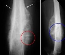
A 12 year old boy presented with localized pain and tenderness in the thigh bone.He has complain of fever.On examination of blood ,ESR was found elevated.Frontal and lateral view of Xray is given in the image.What can be the most probable diagnosis?(Red circle,blue circle and white arrows are made in the X ray to look for important findings for diagnosis)
| A |
Osteosarcoma |
|
| B |
Giant Cell Tumor |
|
| C |
Ewing’s Sarcoma |
|
| D |
Osteoid Osteoma |
Ans:C.)Ewing’s Sarcoma
The Image shows:
- Frontal radiograph and lateral radiographs of the femur demonstrating mottled, osteolytic lesion (blue circle) with poorly marginated edges in the diaphysis of the bone.
- There is sunburst periosteal reaction (red circle) and lamellated periosteal reaction (white arrows)
Ewing’s Sarcoma
- Ewing sarcoma is one of the small, round cell lesions of bone
- Second most common malignant bone tumor in children (after osteosarcoma)
- More common in males than females
- Occurs between the ages of 5-30 years
Location
- Arise in medullary cavity, usually of long bones in the lower extremities
- Most commonly occurs in long bones and pelvis
- Most occur in femur, pelvis, tibia, and humerus
- But they can occur in virtually any bone
- Commonly involves metadiaphysis of long bones
Clinical Findings:
- Most common symptoms are localized pain and swelling
- Additional symptoms/signs may include
- Fever
- Weight loss
- Anemia
- Leukocytosis
- Elevated erythrocyte sedimentation rate
Imaging Findings:
- Most lesions are visible on conventional radiographs
- However, their degree of spread is better evaluated with MRI
Common manifestations on conventional radiography include
- Poorly marginated, lytic, destructive lesion
- Permeative (small holes) or moth-eaten (mottled) appearance
- Rarely, they can be sclerotic
- Soft tissue mass or infiltration is common
- Soft tissue mass may occur without destruction of cortex
- Soft tissue mass may produce saucerization (scalloped depression in cortex)
- Periosteal reaction is common
- Lamellated – onion-skinning due to successive layers of periosteal development
- Sunburst or spiculated – hair-on-end appearance when new bone is laid down perpendicular to cortex along Sharpey’s fibers
- Codman’s triangle – formed between elevated periosteum with central destruction of cortex
- Osteosclerosis may be present secondary to reactive bone formation
Prognosis
- 60-75% five-year survival.
Treatment:
- Systemic chemotherapy is the mainstay of treatment with surgery and/or radiotherapy playing a role depending of the location and size of the tumour.
Most common site of Ewing’s sarcoma ‑
| A | Upper end of tibia | |
| B | Shaft of tibia | |
| C |
Lower end of femur |
|
| D |
Shaft of femur |
Ans. is ‘d’ i.e., Shaft of femur
Commonest site
Ivory osteoma → Frontal sinus
Osteoid osteoma → Tibia
Admantinoma (Ameloblastoma) → Mandible
Chordoma → Sacrum and cervical spine
Osteoclastoma → Lower end of femur
Ewing’s sarcoma → Femur diaphysis > tibia diaphysis
Osteosarcoma → Distal femur (45%), Proximal tibia (25%)
Simple bone cyst → Proximal humerus
Aneurysmal bone cyst → Bones around knee (Femur, tibia)
Osteochondroma → Bones around knee (distal femur > proximal tibia)
Fibrous dysplasia → Proximal femur > Tibia
Enchondroma → Phalanges of hand
Multiple myeloma → Vertebrae
Metastasis → Vertebrae
