EXTERNAL EAR
EXTERNAL EAR
- External ear consists of following structures:
– Pinna/ Auricle
– External auditory canal (or external auditory meatus)
– Tympanic membrane (or eardrum)
EMBRYOLOGY:
- First branchial cleft is the precursor of external auditory canal.
- Around the sixth week of embryonic life, a series of six tubercles appear around the first branchial cleft.
- They progressively coalesce to form the auricle.
- Branchial clefts are ectodermal in origin.
- Pinna is formed at birth.
ANATOMY:
Skin
- Thin with no dermal palillae.
- Closely adherent to underlying cartilage & bony wall.
- Cartilagenous part has subcutaneous tissue -secretes wax.
- Active – collumnar & Quiescent – cuboidal
- Ceruminous glands and hair follicles are limited to cartilagenous parts only.
- Skin over pinna is fixed, Loosely on medial side.
Pinna/Auricle

- Irregularly concave, faces forwards with many eminences and depressions
- Helix
- Crus of helix
- Auricular tubercle (Darwin’s tubercle)
- Antihelix & its 2 cruras
- Triangular fossa
- Scaphoid fossa
- Concha & Cympa concha
- Tragus
- Antitragus
- Intertragic notch
- Lobule of pinna
Cartilagenous framework of auricle
- Single thin plate of elastic fibrocartilage (yellow)
- Continous with the cartilage of external auditory canal (EAC).
- No cartilage in lobule and between tragus and crus of the helix.
- Helix and antihelix are separated by fissura anti-tragohelicinia.
- Medial aspect has Eminentia concha & Eminentia scaphae separated by sulcus anti-helicis transversus
- E. conchae is crossed by a oblique ridge – Ponticulus
Ligaments
- Extrinsic – connects auricle to temporal bone
– Anterior Lig – tragus & spine of helix to root of zygomatic process
– Posterior Lig – post surf. of concha to lat. surf of mastoid process
- Intrinsic – connects individual auricular cartilages
– Strong fibrous band between tragus and helix
– Another band between antihelix and tail of helix
MUSCLES:
| TYPES | MUSCLES | NERVE SUPPLY | BLOOD SUPPLY | ACTION |
|
EXTRINSIC MUSCLES
|
|
|
Post. Auricular artery
|
|
|
INTRINSIC MUSCLES
|
|
|
Post auricular & superficial temporal artery | Minimal change in shape of auricle |
INNERVATION OF AURICLE
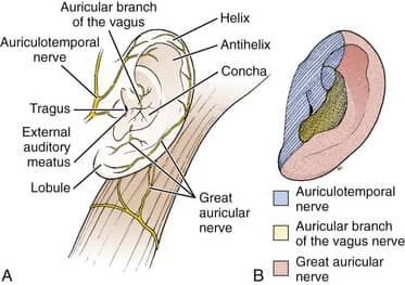
- Great Auricular Nr – most of medial surface & post. part of lateral surface (inclu. lobule)
- Lesser Occipital Nr – upper part of medial surface
- Auriculotemporal Nr – tragus, crus of helix & adjacent helix
- Auricular br of Vagus (Arnold’s Nr) & Facial Nr – Concha (lat.) & Eminentia concha (med.), post. auricular skin
- Pinna is supplied by Vagus nerve, Auriculotemporal nerve, Greater auricular nerve
Exam Important
- Pinna is ectodermal in origin
- Pinna is formed at birth
- Pinna is composed of a thin plate of yellow elastic cartilage, covered with integument
- Pinna develops from the cleft of Ist arch
- Sensory nerve supply of pinna is by V3
- Major part of the skin of pinna is supplied by Great auricular nerve
- Pinna is supplied by Vagus nerve, Auriculotemporal nerve, Greater auricular nerve
- Skin over pinna is fixed Loosely on medial side
Don’t Forget to Solve all the previous Year Question asked on EXTERNAL EAR
Click Here to Start Quiz
Click Here to Start Quiz
