
FALLOPIAN TUBE
| A | Interstitial portion | |
| B |
lsthmus |
|
| C |
lnfundibulum |
|
| D |
Ampulla |
Narrowest part of Fallopian Tube is:
| A |
Interstitial portion |
|
| B |
lsthmus |
|
| C |
lnfundibulum |
|
| D |
Ampulla |
Ans. is B. Isthmus
The fallopian tube is described as having four parts (lateral to medial);
- Fimbriae – finger-like, ciliated projections which capture the ovum from the surface of the ovary.
- Infundibulum – funnel-shaped opening near the ovary to which fimbriae are attached.
- Ampulla – widest section of the uterine tubes. Fertilization usually occurs here.
- Isthmus – narrow section of the uterine tubes connecting the ampulla to the uterine cavity.
| A |
Infundibulum – Ampula – Isthmus – Intramural |
|
| B |
Ampula – Isthmus – intramural – Infundibulum |
|
| C |
Infundibulum – Ampula -Intramural -Isthmus |
|
| D |
Isthmus – Ampula – Infundibulum – Intramural |
Infundibulum – Ampula – Isthmus – Intramural
The length of fallopian tube is :
| A |
8-10 cm |
|
| B |
10 – 12 cm |
|
| C |
15 – 18 cm |
|
| D |
18 – 20 cm |
10 – 12 cm
Longest part of the fallopian tube is :
| A |
Interstitial portion |
|
| B |
Ampulla |
|
| C |
Infundibulum |
|
| D |
Isthmus |
Ampulla
Fallopian tube patency is checked by
| A |
Hysterosalpingography |
|
| B |
Laparoscopy |
|
| C |
Hysteroscopy |
|
| D |
All of the above |
All of the above
Tests Performed for Tubal patency are : 1. CO, insufflation test/ Rubins test (out dated)°
- Hysterosalpingography (HSG)°: Screening test.
- Laparoscopic chromotubation°: Best test
- Sonosalpingography (Sion Test)°
- Hysteroscopy°
- Transcervical Falloscopy°
- Ampullary and Fimbrial Salpingoscopy.°
Extra Edge : Time of performing Tubal Patency test 61‘ – 111h day of cycle (-= 10′h day).
Result :
- If tubes are patent, the medium will be seen to spill out of the abdominal ostia on to the adjacent viscera.°
- If either of the tubes is blocked – site will be shown.°
- A hydrosalpinx will show as a large confined mass of dye without peritoneal spill.°
- Bilateral cornual block with extravasation of the dye is suggestive of tubercular salpingitis.° • Laparoscopic chromotubation -Once HSG shows blocked tubes, it can be confirmed by laparoscopic visualization of the pelvis, fallopian tubes and ovary and injection of methylene blue through the cervix to visualize free spill or absence of spill.
In addition to tuba! patency it can diagnose any peritubal adhesions or endometriosis.
Sonosalpingography -(SION TEST) : In this test, 200 ml of physiological saline is slowly injected into the uterine cavity with the help of foleys catheter (its inflated bulb lies at the level of internal os) and flow of the saline along the tube is then visualized by USG.
| A | Vagina | |
| B |
Vulva |
|
| C |
Ovary |
|
| D |
Fallopian Tubes |
- They are abundant during the premenstrual and menstrual periods.
- They have long slender, rod like nuclei and small amount of cytoplasm squeezed by adjoining cells.
- They represent secretory cells in transitional stages.
Which part of the fallopian tube is the most common site for female tubal sterilization?
| A |
Cornua |
|
| B |
Ampulla |
|
| C |
Isthmus |
|
| D |
Infundibulum |
Most common site for female tubal sterilization is isthmus.
Isthmus is the part of the fallopian tube which is cut or sealed in a tubal ligation or sterilization procedure.
In the bipolar electrocoagulation technique, the mid isthmic portion of the tube is grasped and radiofrequency electric current is applied to three adjacent areas and coagulating 3 cm of tube.
The Hulka clips are also placed across the mid isthmus.
Ref: Maternal and Child Health Nursing: Care of the Childbearing and Childrearing By Adele Pillitteri, page 93
A fertilised ovum reaches the uterine cavity by the 7th day from ovulation. How long does it take for it to travel through the fallopian tube?
| A |
3-4 days |
|
| B |
5-6 days |
|
| C |
7-8 days |
|
| D |
10 days |
The fertilised ovum takes 3-4 days to travel through the fallopian tube and reaches the uterine cavity by the 7th day from ovulation. It is fully embedded in the endometrium by the 14th day.
True about fallopian tubes are all except :
| A |
Lined by cuboidal epithelium |
|
| B |
Isthmus is the narrower part of the tube that links to the uterus |
|
| C |
Tubal ostium is the point where the tubal canal meets the peritoneal cavity |
|
| D |
Mullerian duct develops in females into the Fallopian tubes |
The two uterine tubes are each about 4 in. (10 cm) long and lie in the upper border of the broad ligament.
Each connects the peritoneal cavity in the region of the ovary with the cavity of the uterus.
The uterine tube is divided into four parts:
The infundibulum is the funnel-shaped lateral end that projects beyond the broad ligament and overlies the ovary.
The tubal ostium is the point where the tubal canal meets the peritoneal cavity.
The ampulla is the widest part of the tube.
The isthmus is the narrowest part of the tube and lies just lateral to the uterus.
The intramural part is the segment that pierces the uterine wall.
Function
The uterine tube receives the ovum from the ovary and provides a site where fertilization of the ovum can take place (usually in the ampulla).
The inner mucous membrane of the uterine tube is lined by the ciliated columnar epithelium mixed with the nonciliated secretory cells or peg cells.
The Mullerian ducts develop in females into the fallopian tubes, uterus and vagina, while the Wolffian ducts develop in males into the epididymis and vas deferens
| A |
Isthmus |
|
| B |
Intramural |
|
| C |
Ampulla |
|
| D |
Infundibulum |
Ans. a. Isthmus
Isthmus of the fallopian tube acts as a functional/anatomical sphincter.
The uterine end of the tube, called the isthmus, acts like sphincter, and prevents the embryo from being released into the uterus for 2 days, so that it enters the uterus at the just right time for the implantation.’- How To Have A Baby: Overcoming Infertility By Aniruddha N Malpani/105
‘The lumen of the narrow isthmus is relatively simple, with a few longitudinal folds. This portion of its tube is 2 or 3 cm long. There are three layers of musculature: the inner longitudinal, the middle circular layer, and the outer longitudinal layer. There is some evidence that the isthmus may act as a sphincter.’- Clinical Anatomy of the Uterus, Fallopian Tubes, and Ovaries by Eric R Sokol
| A |
Infundibulum |
|
| B |
Ampulla |
|
| C |
Isthmus |
|
| D |
Interstitial part |
Mucosa of fallopian tube
Mucosa of fallopian tube is folded to form numerous longitudinal folds which project into the lumen. Maximum numbers of these folds are present in the ampulla.
- The mucosa is covered by simple columnar epithelium.
- The epithelium has two types of cells :‑
- Ciliated cells
- Non-ciliated secretory cells (peg cells)
- Secretion of peg cells provides nutrition to ovum.
| A | Interstitial part | |
| B |
Ampulla |
|
| C |
Isthmus |
|
| D |
Fimbria |
Ans. is ‘b’ i.e., Ampulla
The beneficial effects of stay in the female genital tract are collectively called capacitation, from the isthmus, capacitated sperms move rapidly to the ampullas, where fertilization takes place.
Identify the structure as marked in the picture below ?
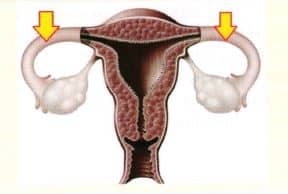
| A |
Fallopian tubes. |
|
| B |
Ovary. |
|
| C |
Cervix. |
|
| D |
None of the above. |
The structure marked in the picture above represents fallopian tubes.
The uterine tube (fallopian tube) carries an egg from the ovary to the uterus. Unless a biological abnormality, surgery, or ectopic pregnancy caused the loss of one tube, women should have two uterine tubes in their bodies.
All are true about the structure marked by an arrow in the picture below except ?
| A |
Lined by cuboidal epithelium. |
|
| B |
Isthmus is the narrower part of the tube that links to the uterus. |
|
| C |
Tubal ostium is the point where the tubal canal meets the peritoneal cavity. |
|
| D |
Mullerian ducts develops in females into the fallopian tubes. |
The structure marked in the picture above represents fallopian tubes.
The two uterine tubes are each about 4 in. (10 cm) long and lie in the upper border of the broad ligament.
Each connects the peritoneal cavity in the region of the ovary with the cavity of the uterus.
The uterine tube is divided into four parts:
The infundibulum is the funnel-shaped lateral end that projects beyond the broad ligament and overlies the ovary.
The tubal ostium is the point where the tubal canal meets the peritoneal cavity.
The ampulla is the widest part of the tube.
The isthmus is the narrowest part of the tube and lies just lateral to the uterus.
The intramural part is the segment that pierces the uterine wall.
Function
The uterine tube receives the ovum from the ovary and provides a site where fertilization of the ovum can take place (usually in the ampulla).
The inner mucous membrane of the uterine tube is lined by the ciliated columnar epithelium mixed with the nonciliated secretory cells or peg cells
The Mullerian ducts develops in females into the fallopian tubes, uterus and vagina, while the Wolffian ducts develops in males into the epididymis and vas deferens.
