Fetal circulation
INTRODUCTION:

- The liver and heart of the fetus receive blood with very high oxygen saturation
Placental circulation consists of independent circulation of blood in two systems:
- Uteroplacental circulation
- Fetoplacental circulation
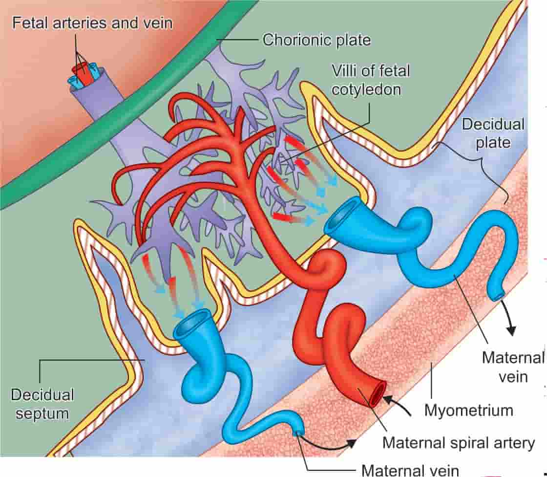
UTEROPLACENTAL CIRCULATION (maternal circulation):
- Circulation of maternal blood through the intervillous space
- The primitive uteroplacental circulation is functionally established during end of first month
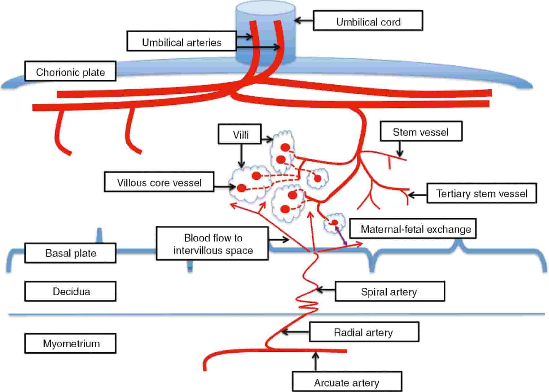
- Volume:500 mL →350 mL in villi system and 150 mL in intervillous space
- Intervillous blood flow at term:500–600 mL per minute, completely replaced about 3–4 times per minute
- Pressure within intervillous space:10–15 mm Hg(uterine relaxation);30–50 mm Hg (uterine contraction)
- Fetal capillary pressure:20–40 mm Hg
Arterial circulation:
- Initailly at 12 weeks cytotrophoblastic invasion into the spiral arteries up to the intradecidual portion
- Secondary invasion of trophoblast between 12 weeks and 16 weeks up to radial arteries
- Spiral arteries are converted to large bore uteroplacental arteries
- Net effect is funneling of the arteries which reduces pressure of blood to 70–80 mm Hg & increases blood flow before it reaches the intervillous space.
- Extravillous trophoblast:Trophoblast cells that do not take part in villous structure
- Endovascular: Invades lumen of the spiral arteries & replaces the endothelium
- Interstitial:Invades inner third of myometrium.
- NK cells prevent Further invasion
Venous drainage:
- Intervillous space → Uterine veins
Circulation in the intervillous space:
- Arterial blood → Chorionic plate → Lateral dispersion → Migration toward the basal plate→ Uterine veins.
FETOPLACENTAL CIRCULATION:
- The two umbilical arteries carry the impure blood from the fetus
- Umbilical arteries → Underneath amnion → Chorionic plate→ Break up → Branches enter stems of the chorionic villi → Primary, secondary and tertiary vessels(villi) → Terminal capillary/shunts → venous channels
- Fetal blood is returned directly to the placenta through the two hypogastric arteries.
- The distal portions of the hypogastric arteries atrophy and obliterate within 3 to 4 days after birth; remnants are called umbilical ligaments.
- Maternal and fetal bloodstreams shows countercurrent flow facilitating material exchange
- Villous capillary pressure:20–40 mm Hg
FETAL BLOOD CHARACHTERISTICS:
- Maximum level of alpha feto protein is seen in Fetal serum
- Heart receives blood with high oxygen saturation
- Blood in IVC has more saturation than blood in SVC as IVC carries the oxygenated blood of umblical vein.
- The left ventricular blood has more oxygen saturation that right ventricular blood because it carries the blood of IVC,while blood in right ventricle is a mixture of blood from IVC and SVC
- Fetal Hb shows high pO2 saturation compared to adult Hb because Affinity to binding to DPG is different in fetal Hb
- The left ventricular output is approximately half of right ventricular output because volume of blood reaching in left atrium is considerably lower than volume of blood reaching in right atrium.
- Aorta and pulmonary trunk are connected by ductus arteriosis, and pulmonary trunk has pressure slightly higher or equal to that of arota –> So, blood flows from pulmonary trunk to aorta
- The pressure in right and left ventricles are equal
- Fetal blood flow:400 mL/min
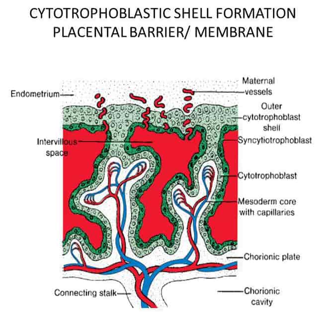
PLACENTAL BARRIER (placental membrane):
In early pregnancy:
- Syncytiotrophoblast
- Cytotrophoblast,
- Basement membrane
- Stromal tissue
- Endothelium of the fetal capillary wall with its basement membrane.
- 0.025 mm thick
Near term:
- Attenuation of syncytial layer
- Sparse cytotrophoblast and distended fetal capillaries almost fill the villus
- Vasculosyncytial membrane: Zone of villi with thin syncytiotrophoblast,(alpha zones for gas exchange)
- Thick “beta zones” of the terminal villi are for hormone synthesis
- IgG passes this barrier
Exam Important
- The primitive uteroplacental circulation is functionally established during end of first month.
- Fetal blood is returned to the umbilical arteries and the placenta through the Hypogastric arteries
- The immunoglobulin which passes the placental barrier in humans is IgG
- Trophoblast, Fetal capillary endothelium & Mesoderm are components of placental barrier
- The liver and heart of the fetus receive blood with very high oxygen saturation
- Maximum level of alpha feto protein is seen in Fetal serum
- Heart receives blood with high oxygen saturation
- Fetal Hb shows high pO2 saturation compared to adult Hb because Affinity to binding to DPG is different in fetal Hb
Don’t Forget to Solve all the previous Year Question asked on Fetal circulation
Click Here to Start Quiz
Click Here to Start Quiz
