GASEOUS EXCHANGE & FACTORS DETERMINING
CO2 diffuses more easily through the respiratory membrane than 02 because it is:
| A |
Less dense |
|
| B |
More soluble in plasma |
|
| C |
Less molecular weight |
|
| D |
Less PCO2 in the alveoli |
CO2 diffuses more easily through the respiratory membrane than 02 because it is:
| A |
Less dense |
|
| B |
More soluble in plasma |
|
| C |
Less molecular weight |
|
| D |
Less PCO2 in the alveoli |
Ans. is. B. More soluble in plasma [Ref: Guyton I 1/e p493]
- Diffusion of gases through the tissues or respiratory membrane depends on the solubility of the gas in lipids; but since all the gases of respiratory importance are all highly soluble in lipids the diffusion through the tissues would depend on diffusion of gases in tissue water.
- The diffusion of gases through fluid depends on multiple factors
(1. the solubility of the gas in the fluid, 2. the cross-sectional area of the fluid, 3. the distance through which the gas must diffuse, 4.the molecular weight of the gas, and 5. the temperature of the fluid.]
of which the two which are characterstics of the gas itself are
– Solubility in fluid &
– Molecular weight.
- The diffusion coefficient of the gas is directly proportional to the solubility and inversely proportional to the square root of the molecular weight.
Diffusion coefficient = Solubility/ Molecular weight
- Diffusion coeffiecient of:
Oxygen → 1.0
Carbon dioxide → 20.3
- Carbon dioxide despite having higher molecular weight than Oxygen, has a very high diffusion coefficient because of its very high solubility in plasma (about 22 times).
| A | Particle’s concentration difference across the membrane | |
| B |
Thickness of the Membrane |
|
| C |
Area of the membrane |
|
| D |
Temperature of the solution |
Which of the following statement regarding COPD is true?
| A |
Decrease FEV1 |
|
| B |
Increased RV |
|
| C |
Diffusion capacity is decreased |
|
| D |
All of the above |
Spirometry findings in COPD includes reduced FEV1 and a reduced FEV1 / FVC ratio. Diffusion capacity for carbon monoxide reflects the ability of lung to transfer gas across alveolar/capillary interface. Diffusion capacity is low in patients with emphysema and infiltrative lung diseases. It is increased in patients with pulmonary hemorrhage, congestive heart failure and asthma.
Reference:
Current Medical Diagnosis and Treatment 2013, chapter 9.
Which of the following does not affect rate of diffusion?
| A |
Area of diffusion |
|
| B |
Concentration gradient |
|
| C |
Time |
|
| D |
None of the above |
The magnitude of the diffusing tendency from one region to another is directly proportionate to the cross-sectional area across which diffusion is taking place and the concentration gradient, or chemical gradient, which is the difference in concentration of the diffusing substance divided by the thickness of the boundary (Fick’s law of diffusion).
Ref:Ganong’s Review of Medical Physiology 23rd edition, Chapter 1.
Gas used to measure diffusion in lung:
| A |
CO |
|
| B |
NO |
|
| C |
CO2 |
|
| D |
Nitrogen |
A i.e. CO
Partition coefficient of gas
| A |
Measure of potency |
|
| B |
Directly proportional to potency |
|
| C |
Measures solubility |
|
| D |
All of the above |
C i.e. Measures solubility
Least diffusion coefficient is for :
| A |
Isoflurane |
|
| B |
Enflurane |
|
| C |
Halothane |
|
| D |
N20 |
D i.e N20
All the following are true about Chronic Obstructive lung disease except:
| A |
Decreased FeV1 |
|
| B |
Decreased MEFR |
|
| C |
Increased RV |
|
| D |
Decreased diffusion capacity |
Answer is D (Decreased diffusing capacity) :
Diffusion capacity is not affected in Obstructive lung diseases 9(acute or chronic).
It is decreased in restrictive lung disease.
- Decreased in expiratory flow rate is a hallmark of obstructive lung disease. (i,MEFR)
- Residual volume is increased & FEVI/FVC is decreased (FEV1 is also decreased).
FEV1=forced expiratory volume in one second; FVC=Forced Vital Capacity; FEF25_75=Forced Expiratory Flow at 25%=75% vital capacity; TLC= Total Lung Capacity; DLCO=Diffusion Capacity of the Lung for Carbon monoxide.
| A |
Emphysema |
|
| B |
Primary pulmonary hypertension |
|
| C |
Alveolar haemorrhage |
|
| D |
Infiltrative lung disease |
Answer is C (Alveolar hemorrhage):
An elevated D Lco is characteristic of alveolar haemorrhage as in Good pastures syndrome.
Hemoglobin contained in erythrocytes in alveolar lumen binds carbon monoxide, so that exhaled CO concentration is diminished and the measured Di increased.
Diffusion capacity
This is measured by estimating Dix() (carbon monoxide diffusion).
It is most useful in assessing disease affecting the
Alveolar capillary bed (interstitial lung disease, emphysema)
Pulmonary vasculature (pulmonary embolism or prim. pul. hypertension)
Decreased Deco (Diffusion capacity)
- Interstitial lung disease: scarring of alveolar capillary units diminishes area of alveolar capillary bed as well as pulmonary blood volume
- Emphysema: alveolar walls are destroyed so the surface area of alveolar capillary bed is diminished
- Recurrent pulmonary embolism and Primary pulmonary hypertension: disease causes a decrease in cross sectional area and volume of pulmonary vasculature
Increased Di,co (Diffusion capacity)
- Alveolar haemorrhage: as in Good pasture’s syndrome: haemoglobin contained in erythrocytes in alveolar lumen binds Co so exhaled carbon monoxide concentration is diminished & Deco is increased
- Congestive heart failure: May be elevated if pulmonary blood volume is increased. Once pulmonary edema ensues DLCO may decrease as and the net Deco depends on the opposing influences.
| A | Chronic Bronchitis | |
| B |
Emphysema |
|
| C |
Interstitial lung disease |
|
| D |
Pulmonary embolism |
Answer is A (Chronic Bronchitis):
Diffusion capacity for carbon monoxide (DLCO) is usually normal in chronic bronchitis and other obstructive lung diseases with the exception of Emphysema where it is decreased.
Diffusion capacity for carbon monoxie (DLCO)
Diffusion capacity is altered in disease affecting the functional integrity of the alveolar capillary membrane.
In Emphysema, alveolar walls are destroyed, so the surface area of alveolar–capillary bed is diminished resulting in diminished DL CO.
Three main categories are associated with lowered DLCO
- Emphysema Q
- Interstitial Lung Disease Q
- Pulmonary vascular disease (Recurrent Pulmonary emboli or Primary Pulmonary Hypertension)Q
| A | Decreased vital capacity | |
| B |
Increased diffusion capacity for carbon monoxide (DLCO) |
|
| C |
Increased Total Lung capacity |
|
| D |
Decreased FEV |
Answer is B (Increased diffusion capacity for carbon monoxide (DLCO)):
Emphysema is associated with a decreased diffusion capacity for carbon monoxide
Lung Function Tests in Emphysema
TLC : Normal or Increased
FEV : Decreased (< 80% predicted) (Decreased out of proportion to FVC)
FVC : Decreased
FEV1 / FVC : Decreased (< 0.7)
FEF25 – 75 : Decreased (
DLCO : Decreased
Note that DLCO is normal in other obstructive lung diseases like chronic bronchitis
Diffusion capacity for carbon monoxie (DLCO)
Diffusion capacity is altered in disease affecting the functional integrity of the alveolar capillary membrane.
In Emphysema, alveolar walls are destroyed, so the surface area of alveolar–capillary bed is diminished resulting in diminished DLCO.
Three main disoders are associated with lowerd DLCO
- Emphyseinae
- Interstitial Lung Diseasee
- Pulmonary vascular disease (Recurrent Pulmonary emboli or Primary Pulmonary Hypertension)Q
| A | Diffusion | |
| B |
Receptor mediated |
|
| C |
Active transport |
|
| D |
Osmosis |
Ans. is ‘a’ i.e., Diffusion
Oxygen is transferred from the alveoli to the pulmonary capillaries by the process of diffusion. The diffusion occurs across the respiratory membrane.
As oxygen diffuses into the blood, it combines with hemoglobin. However, the oxygen, in combination with hemoglobin, does not contribute to the PO2 of blood. Therefore the alveolar – capillary gradient of PO2 is maintained much longer than it would be if hemoglobin were not present. The sequence mey be visualized as follows:-
i) Oxygen diffuses from the alveoli into the plasma due to the PO2 gradient.
ii) To start with, the PO2 in the RBC fluids is the same as in the plasma. As soon as diffusion from the alveoli to plasma raises the PO2 of plasma above that of the RBC fluids, oxygen diffuses from the plasma into the RBC.
iii) As soon as oxygen enters the RBC, it combines with hemoglobin, which mops up oxygen but does not let PO2 rise. The process continues till hemoglobin cannot pick up any more oxygen.
iv) Then the PO2 in the RBC fluids rises to equal that in the plasma and diffusion from the plasma to RBC stops.
v) After that the PO2 in the plasma also rises to equal that in the alveoli, and consequently diffusion from the alveoli to the plasma stops.
Thus, the presence of hemoglobin makes an enormous difference to the amount of oxygen transferred. By mopping up oxygen without letting the PO2 rise, it lets the diffusion of oxygen continue much longer. While the PO2 gradient determines the direction of diffusion of oxygen, it is actually hemoglobin that lays the trap for carrying large amounts of oxygen in the blood.
| A | Decreased vital capacity. | |
| B |
Increased diffusion capacity for carbon monoxide (DLCO). |
|
| C |
Increased Total Lung capacity. |
|
| D | Decreased FEV. |
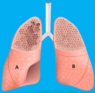
THe lung marked as “A” represent emphysema while the lung marked as “B” is a normal functioning lung.
Emphysema is associated with a decreased diffusion capacity for carbon monoxide
Lung Function Tests in Emphysema
TLC : Normal or Increased
FEV : Decreased (< 80% predicted) (Decreased out of proportion to FVC)
FVC : Decreased
FEV1 / FVC : Decreased (< 0.7)
FEF25 – 75 : Decreased (
DLCO : Decreased
Note that DLCO is normal in other obstructive lung diseases like chronic bronchitis
Diffusion capacity for carbon monoxie (DLCO)
Diffusion capacity is altered in disease affecting the functional integrity of the alveolar capillary membrane.
In Emphysema, alveolar walls are destroyed, so the surface area of alveolar–capillary bed is diminished resulting in diminished DLCO.
Three main disoders are associated with lowerd DLCO
- Emphyseinae
- Interstitial Lung Diseasee
- Pulmonary vascular disease (Recurrent Pulmonary emboli or Primary Pulmonary Hypertension)Q
| A | Inerstitial lung diseas | |
| B |
Goodpasture’s syndrome |
|
| C |
Pneumocystis Jiroveci |
|
| D |
Primary pulmonary hypertension |
Ans. is ‘b’ i.e., Goodpasture’s syndrome
Gas diffusion tests :
- Gas diffusion tests measure the amount of oxygen and other gases that cross the alveoli into the blood.
- These tests evaluate how well gases are being absorbed into the blood from lungs. Gas diffusion tests include.
- Carbon monoxide diffusing capacity (transfer factor DLcy)
- Arterial blood gases
Carbon monoxide diffusing capacity (DL):
- This measures how well the lung transfers a small amount of carbon monoxide into the blood0.
- Normally, in the lung, a gas has to cross the alveolar membrane, capillary membrane to reach the blood where it combines with hemoglobin.
So quiet obviously the diffusion capacity of gas depends upon
- Driving pressure of the gas
- Surface area of alveolar capillary membrane
- Thickness of alveolar capillary membrane
- Diffusion coefficient of the gas
- Red blood cell volume.
- Reaction rate with hemoglobin and hemoglobin level of patient.
- Degree of V/Q mismatching.