GLAUCOMA
GLAUCOMA

ETIOLOGY:
- It’s the result of high fluid pressure inside eye.
- This happens when the liquid in the front part of the eye doesn’t circulate the way it should.
- Most common etiolopathogenetic cause of glaucoma is Decreased outflow
- Inverse glaucoma occurs in Spherophakia
- Normally, the fluid, called aqueous humor, flows out of eye through a mesh-like channel.
- If this channel gets blocked, the liquid builds up. That’s what causes glaucoma.
- In haemolytic glaucoma the mechanisms are Siderosis of trabeculae,Deposition of haemosiderin & RBC clogging the trabeculae
- The reason for the blockage is unknown, but it can be inherited.
- Less common causes include a blunt or chemical injury to eye, severe eye infection, blocked blood vessels inside the eye, and inflammatory conditions.
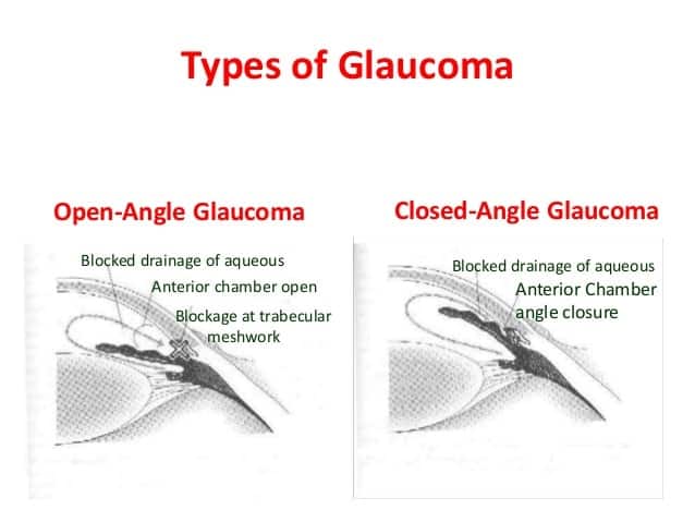
TYPES:
There are two main kinds:
Open-angle glaucoma:
- It’s the most common type.
- First sign seen in open-angle glaucoma is Extension above blind spot
- The drain structure in eye , it’s called the trabecular meshwork
- Open anterior chamber angle
- Frequent change in presbyopic correction
- Looks normal, but fluid doesn’t flow out like it should.
-
Also known as chronic simple glaucoma of adult onset and is typically characterized by slowly progressive raised intraocular pressure.
Associated with characteristic optic disc cupping and specific visual field defects.
Pathogenesis-
Heredity: POAG has a polygenic inheritance.
Age- elderly between 5th and 7th decades.
Myopes are more predisposed than the normals.
Diabetics have a higher prevalence of POAG.
POAG is more in hyPertensives.
C/F
patients usually complain of frequent changes in presbyopic glasses.
Patients develop delayed dark adaptation.
Fundus examination show large cup. (0.6 or more) -
Laser iridotomy is used for treatment of primary angle closure glaucoma
Variations of OAG include:
- Primary open angle glaucoma (POAG),
- Normal-tension glaucoma (NTG),
- Pigmentary glaucoma,
- Pseudoexfoliation glaucoma,
- Secondary glaucoma Congenital glaucoma(presents as Photophobia)
-
-
- seen with Diffuse iris melanoma
- Secondary glaucoma following corneal perforation is due to Peripheral anterior synechiae
-
-
Angle-closure glaucoma:
- Also called acute or chronic angle-closure or narrow-angle glaucoma.
- Shallower Anterior chamber & Short axial length of eyeball (Hypermetropia)
- Eye doesn’t drain right because the angle between iris and cornea is too narrow & iris is in the way.
- This can cause a sudden buildup of pressure in eye.
- It’s also linked to farsightedness and cataracts, a clouding of the lens inside eye.
- Variations of narrow angle glaucoma include
- Acute angle closure glaucoma,
- Chronic angle closure glaucoma
-
-
- Pupillary block: uveitis, psudophakia
- Angle fibrosis: neovascular glaucoma
-
-
Neovascular glaucoma
- Most commonly due to diabetes & CRVO
- Other:CRAO,Carotid artery obstructive disease, Rhegmatogenous retinal detachment,Choroidal melanoma, Sickle-cell retinopath, Carotid-cavernous fistula
SYMPTOMS:
- The earliest change in glaucoma is Baring of the blind spot
- Glaucoma often is called the “silent thief of sight,” because most types typically cause no pain and produce no symptoms until noticeable vision loss occurs.
- Pupil in acute congestive glaucoma Vertically oval & semidilated
- In chronic simple glaucoma the most common field defect is Baring of blind spot
- Patient with open angle glaucoma of myopia, complains of blurring of vision on administration of pilocarpine due to Small pupil
- For this reason, glaucoma often progresses undetected until the optic nerve already has been irreversibly damaged, with varying degrees of permanent vision loss.
- Hundred day glaucoma is associated with Central retinal vein occlusion(CRVO)
- Tears of the iris sphincter and ciliary body is seen in angle recession glaucoma with Angle recession more than 180 degree
- Secondary open angle glaucoma can occur in angle recession glaucoma
With acute angle-closure glaucoma, symptoms that occur suddenly can include :
- Blurry vision,
- Halos around lights,
- Intense eye pain,
- Nausea and vomiting.
- If you have these symptoms, make sure you see an eye care practitioner or visit the emergency room immediately so steps can be taken to prevent permanent vision loss.
- Double arcuate or ring shaped scotoma in glucoma develops when two arcuate scotomas join.
DIAGNOSIS & TEST:
- The most reliable provocative test for angle-closure glaucoma is Dark room test
- Tonometer used to measure your intraocular pressure
- Sophisticated imaging technology to create baseline images and measurements of the eye’s optic nerve and internal structures.— such as Visual field testing is a way for your eye doctor to determine if you are experiencing vision loss from glaucoma.
- Scanning laser polarimetry (SLP),
- Optical coherence tomography (OCT)
- Confocal scanning laser ophthalmoscopy
- Gonioscopy also may be performed to make sure the aqueous humor (or “aqueous”) can drain freely from the eye.
Exam Important
- Double arcuate or ring shaped scotoma in glucoma develops when two arcuate scotomas join.
- Most common cause of neovascular glaucoma Diabetes
- Pupil in acute congestive glaucoma Vertically oval & semidilated
- Patient with open angle glaucoma of myopia, complains of blurring of vision on administration of pilocarpine due to Small pupil
- Pseudophakia is a cause of secondary angle closure glaucoma
- Hundred day glaucoma is associated with CRVO
- Neovascular glaucoma is caused by CRVO, CRAO, Diabetes mellitus
- Tears of the iris sphincter and ciliary body is seen in angle recession glaucoma
- Angle recession more than 180 degree in angle recession glaucoma
- Secondary open angle glaucoma can occur in angle recession glaucoma
- Frequent change in presbyopic correction is seen in open angle glaucoma
- open angle glaucoma is most common with Open anterior chamber angle
- In angle closure glaucoma Small cornea, shallower Anterior chamber & Short axial length of eyeball are the anatomical changes seen
- Angle closure glaucoma may be associated with Hypermetropia
- Most common etiolopathogenetic cause of glaucoma is Decreased outflow
- Intractable secondary glaucoma is seen in Diffuse iris melanoma
- Congenital glaucoma presents as Photophobia
- First sign seen in open-angle glaucoma is Extension above blind spot
- The most reliable provocative test for angle-closure glaucoma is Dark room test
- In haemolytic glaucoma the mechanisms are Siderosis of trabeculae,Deposition of haemosiderin & RBC clogging the trabecular
- Secondary glaucoma following corneal perforation is due to Peripheral anterior synechiae
- The earliest change in glaucoma is Baring of the blind spot
- In chronic simple glaucoma the most common field defect is Baring of blind spot
- Inverse glaucoma occurs in Spherophakia
Don’t Forget to Solve all the previous Year Question asked on GLAUCOMA
Click Here to Start Quiz
Click Here to Start Quiz
