Healing of Skin wounds
| A | End of first week | |
| B |
End of second week |
|
| C |
End of third week |
|
| D |
End of 2 months |
Maximum collagen in wound healing is seen at –
| A |
End of first week |
|
| B |
End of second week |
|
| C |
End of third week |
|
| D |
End of 2 months |
Ans. is ‘c’ i.e., End of the third week
|
Day
|
Features of wound
|
|
Day 0 (when the wound has formed)
|
Presence of blood clot in the incision
|
|
Day 1 (within 24 hours)
|
a neutrophilic infiltration blood clot
|
|
Day 2 (24 to 48 hours)
|
neutrophils blood clot continuous thin epithelial layer
|
|
Day 3
|
Macrophages replace neutrophils, Appearance of granulation tissue, type III collagen deposition begins but do not bridge the incision
|
|
Day 5
|
Abundant granulation tissue
collagen fibrils bridge the incision
|
|
End of 2nd week
|
accumulation of collagen fibroblast proliferation
|
|
1 month
|
Replacement of collagen type III with collagen type I (has greater tensile strength) due to action of collagenase enzyme
|
| A |
Thin continuous epithelial cover appears |
|
| B |
Fibroblasts lay down collagen fiber |
|
| C |
Granulation tissue fills the wound |
|
| D |
Neutrophils line the wound edge |
Ans. is ‘d’ i.e., Neutrophils line the wound edge
Wound healing is the summation of following processes except –
| A |
Coagulation |
|
| B |
Matrix synthesis |
|
| C |
Angiogenesis |
|
| D |
Fibrolysis |
Ans. is ‘d’ i.e., Fibrolysis
Primary intentional healing which is true ‑
| A |
Neovascularization is maximum by day 5 |
|
| B |
Neovascularization is maximum by day 3 |
|
| C |
Neutrophils appear at wound margins on day 3 |
|
| D |
The epidermis recovers its maximum thickness by day 7 |
Ans. is ‘a’ i.e., Neovascularization is maximum by day 5
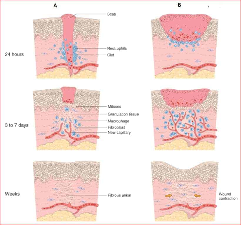
Skin wound healing
- Skin wounds are classically described to heal by primary or secondary intention.
A. Healing by primary intention
- It occurs in wounds with opposed edges, e.g., surgical incision.
- The healing process follows a series of sequantial steps : ‑
Immediate after incision
- Incisional space filled with blood containing .fibrin and blood cells.
- Dehydration of the surface clot forms scab that covers the wound.
Within 24 hours
- Neutrophils appear at the margins of wound.
In 24-48 hours
- Epithelial cells move from the wound edges along the cut margin of dermis, depositing basement membrane components as they move.
- They fuse in the midline beneath the surface scab, producing a continuous but thin epithelium layer that closes the wound.
By day 3
- Neutrophils are largely replaced by macrophages.
- Granulation tissue progressively invades the incision space.
- Collegen fibers now present in the margin but do not bridge the incision.
By day 5
- Incisional space is largely filled with granulation tissue.
- Neovascularization is maximum.
- Collegen fibrils become more abundant and begin to bridge the incision.
- The epidermis recovers its normal thickness.
During second week
- Leukocytes and edema have disappeared.
- There is continued accumulation of collegen and proliferation of fibroblast.
- By the end offirst month
- Scar is made up of a cellular connective tissue devoid of inflammatory infiltrate covered now by intact epidermis.
B. Healing by secondary intention
- It occurs in wounds with seperated edges in which there is more extensive loss of cells and tissue.
- Regeneration of parenchymal cells cannot completely restore the original architecture, and hence abundant granulation tissue grows.
Healingby secondary from primary intention in several respects :
- Inflammatory reaction is more intense.
- Much larger amounts of granulation tissue are formed.
- Wound contraction occurs → Feature that most clearly differentiate secondary from primary healing.
- Permanent wound contraction requires the action of myolifibroblasts – Fibroblasts that have the ultrastructural characteristic of smooth muscle cells.
| A | Wound is clean | |
| B |
Scanty granulation tissue |
|
| C |
Sutures are not used |
|
| D |
Outcome is neat linear scar |
Ans:C.)Sutures are not used
Image shows:’A’:Healing by primary intention,’B’:Healing by secondary intention.
