
HYDATID CYST OF LIVER
HYDATID CYST OF THE LIVER
- Hydatid cyst occurs as a result of infection by the larval cyst stage of the tapeworm, Echinococcus granulosus.
- Dog is the common definite host.
- Intermediate host is sheep, man.

Life cycle of Hydatid Cyst-

ETIOLOGY-
- MC organism – Echinococcus granulosus (unilocular cyst)
- E. Multilocularis- multilocular cyst and can cause malignant hydatidosis.
MORPHOLOGICAL FEATURES-
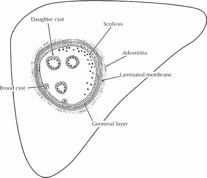
- Cyst wall has 3 layers-
a) Pericyst (adventitia/ pseudocyst)
- Outer host inflammatory reaction.
- Fibrous layer
b) Ectocyst (laminated membrane)
- Intermediate layer. White and elastic.
c) Endocyst-
- Innermost germinal layer
- Secrets hydatid fluid inside.
- Hydatid sand- grain like material composed of scolices present in fluid.

CLINICAL FEATURES-
- Route of infection- faeco- oral route
- Equal in male and female
- Organs involved- liver> lungs> spleen > kidney > brain > bone
- Pain in upper abdomen
- Hepatomegaly
- Typical hydatid thrill- by 3 finger method
- Asymptomatic
- Liver cysts occur more frequently in the right lobe.
- Least common site of calcified hydatid cyst.
INVESTIGATIONS-
- USG- detect localise and used for aspiration of cyst
- X- ray abdomen- speckled calcification
- CT scan
- ERCP- drainage of hydatid contents into duodenum
- Casoni’s intradermal cyst- sensitivity and specificity test is low. Not in use now.
- ELISA and immunoelectrophoresis- shows diagnosis (Serological tests are the most sensitive and specific tests; includes Immunoblast test, arcs test, ELISA and indirect hemagglutination tests)

TREATMENT-
- Albendazole- 400 mg BD for 21 days
- Surgery-
- Laprotomy and isolation of the cyst from peritoneal cavity in hypochlorite solution
- Aspirate and Inject scolicidal agents .
- Incise the cyst
- Percutaneous drainage-
- Also called as PAIR (Puncture, aspiration, injection and reaspiration (MC technique)
- Cyst evacuation + omentopexy/ omentoplasty
- Pericystectomy- most effective treatment for hydatid cyst
- Hepatic resection- kost radial treatment for hydatid cyst
- SCOLICIDAL AGENTS-
a) Hypertonic saline (20%)
b) 0.5% cetrimide with 0.05% chlorhexidine
c) Absolute alcohol
d) 10% iodine
INDICATIONS FOR SURGERY-
- Unilocular uncomplicated cyst
- Poor surgical candidate
- Previous multiple abdominal surgeries
- Relapse following surgery
CONTRAINDICATIONS FOR SURGERY-
- Multiple septal divisions
- Communicating cysts
- Dead or inactive cysts
- Laparoscopic approach
Exam Important
- Hydatid cyst occurs as a result of infection by the larval cyst stage of the tapeworm, Echinococcus granulosus.
- Dog is the common definite host.
- Intermediate host is sheep, man.
MORPHOLOGICAL FEATURES-
- Cyst wall has 3 layers-
a) Pericyst (adventitia/ pseudocyst)
- Outer host inflammatory reaction.
- Fibrous layer
b) Ectocyst (laminated membrane)
- Intermediate layer. White and elastic.
c) Endocyst-
- Innermost germinal layer
- Secrets hydatid fluid inside.
- Hydatid sand- grain like material composed of scolices present in fluid.
CLINICAL FEATURES-
- Route of infection- faeco- oral route
- Equal in male and female
- Organs involved- liver> lungs> spleen > kidney > brain > bone
- Asymptomatic
- Liver cysts occur more frequently in the right lobe.
- Least common site of calcified hydatid cyst.
INVESTIGATIONS-
- USG- detect localise and used for aspiration of cyst
- X- ray abdomen- speckled calcification
- CT scan
- ERCP- drainage of hydatid contents into duodenum
- Casoni’s intradermal cyst- sensitivity and specificity test is low. Not in use now.
- ELISA and immunoelectrophoresis- shows diagnosis (Serological tests are the most sensitive and specific tests; includes Immunoblast test, arcs test, ELISA and indirect hemagglutination tests)
INVESTIGATIONS-
- USG- detect localise and used for aspiration of cyst
- X- ray abdomen- speckled calcification
- CT scan
- ERCP- drainage of hydatid contents into duodenum
- Casoni’s intradermal cyst- sensitivity and specificity test is low. Not in use now.
- ELISA and immunoelectrophoresis- shows diagnosis (Serological tests are the most sensitive and specific tests; includes Immunoblast test, arcs test, ELISA and indirect hemagglutination tests)
TREATMENT-
- Albendazole- 400 mg BD for 21 days
- Surgery-
- Laprotomy and isolation of the cyst from peritoneal cavity in hypochlorite solution
- Aspirate and Inject scolicidal agents .
- Incise the cyst
- Percutaneous drainage-
- Also called as PAIR (Puncture, aspiration, injection and reaspiration (MC technique)
- Cyst evacuation + omentopexy/ omentoplasty
- Pericystectomy- most effective treatment for hydatid cyst
- Hepatic resection- kost radial treatment for hydatid cyst
- SCOLICIDAL AGENTS-
a) Hypertonic saline (20%)
b) 0.5% cetrimide with 0.05% chlorhexidine
c) Absolute alcohol
d) 10% iodine
INDICATIONS FOR SURGERY-
- Unilocular uncomplicated cyst
- Poor surgical candidate
- Previous multiple abdominal surgeries
- Relapse following surgery
CONTRAINDICATIONS FOR SURGERY-
- Multiple septal divisions
- Communicating cysts
- Dead or inactive cysts
- Laparoscopic approach
Don’t Forget to Solve all the previous Year Question asked on HYDATID CYST OF LIVER
Click Here to Start Quiz
Click Here to Start Quiz

