
INGUINAL CANAL
True about boundaries of inguinal canal are all EXCEPT:
| A | Conjoint tendon anteriorly | |
| B |
Fascia transversalis is posterior boundary |
|
| C | Inguinal ligament forms base | |
| D |
Internal oblique forms roof |
True about boundaries of inguinal canal are all EXCEPT:
| A |
Conjoint tendon anteriorly |
|
| B |
Fascia transversalis is posterior boundary |
|
| C |
Inguinal ligament forms base |
|
| D |
Internal oblique forms roof |
Ans.A. Conjoint tendon anteriorly
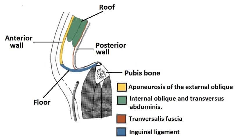
To help define the boundaries, the canal is often further approximated as a box with six sides. Not including the two rings, the remaining four sides are usually called the “anterior wall”, “posterior wall”, “roof”, and “floor”
These consist of the following:
M – Muscles
A – Aponeuroses (The A in MALT coincides with the position of the wall—anterior. This can be used to remember that the direction of the mnemonic is anticlockwise)
L – Ligaments
T – Transversalis/Tendon
| A |
Interfoveolar ligament |
|
| B |
Parietal peritoneum |
|
| C |
Fascia transversalis |
|
| D |
Internal oblique |
Internal oblique
Which of the following structures pass behind the inguinal ligament?
1. Femoral branch of genitofemoral nerve
2. Superficial epigastric artery
3. Psoas major
4. Femoral vein
5. Saphenous vein
| A |
1,2 & 3 |
|
| B |
3 & 4 |
|
| C |
1,3,4 & 5 |
|
| D |
All |
Ans.B
Three small cutaneous arteries (superficial external pudendal, superficial epigastric and superficial circumflex iliac arteries) arising from the femoral artery can be seen a little below the inguinal ligaments.
The femoral branch of the genitofemoral nerve pierces the femoral sheath and overlying deep fascia 2 cm below the mid inguinal point.
The great saphenous vein pierces the saphenous opening in the deep fascia (fascia lata) of the thigh and joins the femoral vein 4 cm below and lateral to the pubic tubercle.
So from the above discussion, it is clear that the femoral branch of the genitofemoral nerve, superficial epigastric artery and saphenous vein passes below (not behind) the inguinal ligament.
Psoas major insertion: The muscle passes behind the inguinal ligament and in front of the hip joint to enter the thigh.
The femoral vein leaves the thigh by passing behind the inguinal ligament.
All are TRUE about the relations of inguinal canal, EXCEPT
| A |
Internal oblique forms the roof |
|
| B |
Inguinal ligament forms the floor |
|
| C |
Fascia transversalis forms posterior wall |
|
| D |
Interfoveolar ligament forms lateral two third of anterior wall |
Ans.D
Boundaries of the Inguinal canal:
- Floor is formed by the grooved upper surface of the inguinal ligament
- Roof is formed by the arching lower fibers of Internal oblique and transverse abdominal muscles.
- Anterior wall is formed by the external oblique aponeurosis all through it is extent reinforced laterally by the Internal oblique muscle.
- Posterior wall is formed by the transversalis fascia all through its extent, reinforced medially by the conjoint tendon also known as inguinal falx (Fused fibers of transversus abdominis and internal oblique muscles).
- Spermatic cord
- Ilioinguinal nerve
- Cremaster and its blood supply
- Genital branch of genitofemoral nerve
- Remnants of processes vaginals
- Round ligament of uterus, (remnant of Gubernaculum)
- Genital branch of genitofemoral nerve
- Ilioinguinal nerve
- Cremaster and its blood supply
- Remnants of processes vaginalis.
| A |
Conjoint tendon is seen anteriorly |
|
| B |
Fascia transversalis forms posterior boundary |
|
| C |
Inguinal ligament forms the base |
|
| D |
Internal oblique forms the roof |
Conjoint tendon is formed by the condensation of fibers of internal oblique and transverse abdominis. It forms the posterior wall of inguinal canal.
Boundaries of inguinal canal:
- Anterior wall of inguinal canal is made up of external oblique aponeurosis with some fibers of internal oblique which attach to lateral part of inguinal ligament.
- Posterior wall is formed from transversalis facia, except medially where the conjoint tendon from transversus abdominis intervenes.
- Inferior wall (floor) is formed from the inguinal ligament as it turns inward, and it is composed medially of lacunar ligament.
- Roof consists of lowermost fibers of internal oblique and transversus abdominis as they arch over the canal to join together as conjoined tendon.
| A |
Inguinal ligament forms the floor of inguinal canal |
|
| B |
Internal oblique forms both anterior wall and roof |
|
| C |
Deep inguinal ring is an opening in transversalis abdominis |
|
| D |
Superficial inguinal ring is found in external oblique aponeurosis |
Deep inguinal ring is an oval opening in the fascia transversalis, that lies about ½ inch above the inguinal ligament midway between the anterior superior iliac spine and pubis symphysis. Medial to it lies the inferior epigastric vessels.
Superficial inguinal ring is a triangular gap in the external oblique aponeurosis, which lies above and lateral to the pubic tubercle.
The boundaries of the inguinal triangle include all of the following, EXCEPT:
| A |
Lateral border of rectus abdominis |
|
| B |
Inguinal ligament |
|
| C |
Inferior epigastric artery |
|
| D |
Linea alba |
Inguinal triangle is a region defined by the following structures:
- Rectus abdominis muscle (medially)
- Inguinal ligament (inferiorly)
- Inferior epigastric vessels (superior and laterally)
All pass through deep inguinal ring, EXCEPT?
| A |
Spermatic cord |
|
| B |
Internal spermatic fascia |
|
| C |
Round ligament |
|
| D |
Ilioinguinal nerve |
It transmits the spermatic cord in the male and the round ligament of the uterus in the female into the inguinal canal. Internal spermatic fascia is present in the spermatic cord. Ilio inguinal nerve passes only through the superficial inguinal canal and not through deep.
Inguinal canal: contains the spermatic cord and the ilioinguinal nerve in the male, and the round ligament of the uterus and the ilioinguinal nerve in the female. It is an oblique canal of about 4 cm. long, slanting downward and medialward, and placed parallel with and a little above the inguinal ligament; it extends from the deep inguinal ring laterally to the superficial inguinal ring medially.
- Spermatic cord in males
- Round ligament in females
- Spermatic cord in males
- Round ligament in females
- Ilioinguinal nerve in both males and females
| A |
Interfoveolar ligament |
|
| B |
Parietal peritoneum |
|
| C |
Fascia transversalis |
|
| D |
Internal oblique muscle |
- Along the whole length: fascia transversalis, extraperitoneal tissue, parietal peritoneum, inferior epigastric vessels.
- Along the medial two-thirds of the canal: Conjoint tendon, reflected part of inguinal ligament and interfoveolar ligament in the lateral one third.
- Along the whole length of canal: skin, superficial fascia, external oblique aponeurosis.
- Along the lateral third of the canal: The fleshy fibers of the internal oblique muscle are also present deep to the external oblique aponeurosis.
True about inguinal canal:
| A |
It is an Intermuscular canal |
|
| B |
Superficial inguinal ring is situated superior and lateral to pubic tubercles |
|
| C |
In male vas deference passes through it. |
|
| D |
All |
A. i.e. It is an intermuscular canal; B. i.e. Superficial inguinal ring is situated superior & lateral to pubic tubercles; D. i.e. In males vas deferens passes through it
All are true about inguinal canal except :
| A |
Conjoint tendon forms part of the posterior wall |
|
| B |
Superficial ring is found in external oblique aponeurosis |
|
| C |
Deep ring is an opening in transversalis abdominis |
|
| D |
Internal oblique forms both anterior and posterior wall |
Ans: C i.e. Deep inguinal ring is an opening in transversalis abdominis
The deep inguinal ring is an opening in the transversalis fascia, not in the transverse abdominis.
Anatomy of the inguinal canal:
It is a 4 cm long intermuscular canal directed downwards, forwards and medially.
It extends from deep inguinal ring to superficial ring.
Boundaries:
Anterior:
- External oblique aponeurosis
- Internal oblique muscle (in lateral 1/3rd)
Superior wall/roof:
- Internal oblique
- Transverse abdominis
Posterior wall:
- Fascia transversalis
- Conjoint tendon (Internal oblique + Transverse abdominis)
Inferior wall/floor:
- Inguinal ligament/Poupart ligament (Rolled out Inferior edge of the external oblique aponeurosis and extends from the anterior superior iliac spine to the pubic tubercle)
- Lacunar ligament at medial end (fan-shaped medial expansion of the inguinal ligament)

Deep inguinal ring: Oval opening in fascia transversalis situated 1.25cm above mid-inguinal point.
Superficial inguinal ring: Triangular opening in external oblique aponeurosis situated 1cm above and lateral to pubic crest.
The inguinal canal is NOT bounded posteriorly by:
| A |
Transversalis fascia |
|
| B |
Internal oblique tendon |
|
| C |
Conjoint tendon |
|
| D |
Lacunar ligament |
D i.e. Lacunar ligament
All of the following structures pass through the inguinal canal in females, Except:
| A |
Ilioinguinal nerve |
|
| B |
Round ligament of uterus |
|
| C |
Lymphatics from the uterus |
|
| D |
Inferior Epigastric Artery |
D i.e. Inferior Epigastric Artery
- Inguinal canal is an oblique intermuscular canal in anterior abdominal wall from deep to superficial inguinal rings. It contains (passes) spermatic cord in males, round ligament in females and ilioinguinal nerve in both sexes Constituents of spermatic cord include vas (ductus) deferens, artery of ductus deferens with autonomic sympathetic nerves around it; testicular artery, lymphatics and vein (pampiniform plexus); cremasteric artery & genital branch of genitofemoral nerve supplying cremasteric muscle; and remains of processes vaginalis.
- It contains blood vessels, nerves & lymphatics. The lymphatics drain the uterine region around the entery of uterine tube to the superficial inguinal lymph nodes.
- Genital branch of genitofemoral nerve enters the inguinal canal by deep ring and supplies cremaster muscle and skin of scrotum in males. In females, it accompanies the round ligament and ends in the skin of mons pubis & labium majus(sensory supply)
- Superficial inguinal ring is a triangular hiatus (opening) in external oblique aponeurosis, just above & lateral (i.e. suprolateral) to pubic crest (Gray’s)/ to pubicle tubercleQ (Moore).
- Inferior epigastric vessels neither pass through deep or superficial inguinal rings nor does it travel through inguinal canalQ. However, inferior epigastric vessels are important posterior relations of medial end of inguinal canal. They lie on fascia transversalis and ascend obliquely behind conjoint tendon into posterior portion of rectus sheath.
- Deep inguinal ring is an opening in fascia transversalis
- Lacunar ligament along with inguinal ligament forms inferior wall
- Triangle of Doom is bounded by gonadal vessels (lateral), vas deferens (medial), and peritoneal reflection (base).
| A |
Femoral branch of genitofemoral nerve |
|
| B |
Femoral vein |
|
| C |
Psoas major |
|
| D |
All |
A i.e. Femoral branch of genitofemoral nerve; B i.e. Femoral vein ; C i.e. Psoas major
The testis descends the inguinal canal during…month.
| A |
5th |
|
| B |
6th |
|
| C |
7th |
|
| D |
8th |
C i.e. 7th
Superficial inguinal ring in the female transmits:
| A |
Broad ligament of uterus |
|
| B |
Round ligament of the uterus |
|
| C |
Cardinal ligament |
|
| D |
None of the above |
The superficial inguinal ring is a triangular aperture in the aponeurosis of the external oblique muscle and is situated above and medial to the pubic tubercle.
In the female, the superficial inguinal ring is smaller and difficult to palpate; it transmits the round ligament of the uterus.
The Following Figure Showing the Saggital section of Inguinal Canal . What are the structures forming the Roof of the Canal

| A |
Aponeurosis of External Oblique |
|
| B |
Transverse Abdominal Muscle |
|
| C |
Both of the Above |
|
| D |
None of the Above |
All are contents of inguinal canal except ‑
| A |
Femoral branch of genitofemoral nerve |
|
| B |
Ilioinguinal nerve |
|
| C |
Round ligament of uterus |
|
| D |
Spermatic cord |
Contents of inguinal canal
- Spermatic cord in male or round ligament of uterus in female.
- Ilioinguinal nerve : It enters through the interval between external and internal oblique muscles.
Note: Genital branch of genitofemoral nerve is a component of spermatic cord (not femoral branch of genitofemoral nerve).
Nerve entering the inguinal canal through deep inguinal ring ‑
| A | Ilioinguinal nerve | |
| B |
Pudendal nerve |
|
| C |
Genital branch of genitofemoral |
|
| D |
Superior rectal nerve |
The spermatic cord in males and round ligament of uterus in females, enter the inguinal canal through the deep inguinal ring and pass out through superficial inguinal ring.
Thus consituents of spermatic cord are also components of inguinal canal; these are ductus deferens (vas deferens), testicular artery, cremestric artery, artery to ductus deference, pampiniform plexus, lymphatics, sympathetic plexus, genital branch of genitofemoral nerve, remains of process vaginalis.
Note: Ili oiguinal nerve enters inguinal canal through interval between external and internal oblique muscles (not through deep inguinal ring).
Which is not a content of inguinal canal ‑
| A |
Spermatic cord |
|
| B |
Ilioinguinal nerve |
|
| C |
Genital branch of genitofemoral nerve |
|
| D |
Inferior epigastric artery |
Ans. is ‘d’ i.e., Inferior epigastric artery
Contents of inguinal canal
- Spermatic cord in male or round ligament of uterus in female.
- Ilioinguinal nerve : It enters through the interval between external and internal oblique muscles.
- Genital branch of genitofemoral nerve is a constituent of spermatic cord.
