
LEUKOPLAKIA
LEUKOPLAKIA
- Leukoplakia is described as any white patch or plaque that cannot be rubbed off or characterised clinically or pathologically as another lesion.
- It is the most common premalignant lesion.

ETIOLOGY-
- 6 S
- Smoking
- Spirit (alcohol)
- Spices (tobacco), betel nut
- Sepsis
- Sharp tooth
- Syphilis
- Oral candidiasis (candida albicans)

PATHOLOGY
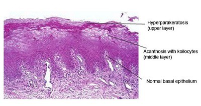
- Microscopic features are-
- Hyperkeratosis- milky blush on the surface
- Acanthosis- elongation of rete pegs (smooth dry patch)
- Parakeratosis- cell are still immature
- Dyskeratosis- forms keratin cell layers in epidermis
- Carcinoma in situ- epithelial pearls seen in dermis.


CLINICAL FORMS OF LEUKOPLAKIA-
- Homogenous
- Non homogenous
- Proliferative verrucous
- Erythroplakia
- Sublingual keratosis
- Oral hairy leukoplakia (Epbstein- Barr virus)
- Syphilitic leukoplakia

CLINICAL FEATURES-
- Seen in middle aged and elderly person
- More common in males.
- Sites- Buccal mucosa, angle of mouth, alveolar mucosa, tongue, lip, hard & soft palate and floor of the mouth.
HISTOLOGY-
- Lesion appears white, whitish- yellow, red- velvety
- Usually are circumscribed, slightly elevated, smooth and wrinkled, speckled or nodular.
- Histological features are hyperkeratosis, dysplasia & anaplasia with nuclear & cellular pleomorphism.
INVESTIGATION-
- Biopsy to rule out malignancy
TREATMENT-
- Severe dysplasia or carcinoma in situ is excised surgically or CO2 laser is used.
- Leukoplakia in vulva treated by Simple vulvectomy; and Estrogen cream.
Exam Important
- Leukoplakia is described as any white patch or plaque that cannot be rubbed off or characterised clinically or pathologically as another lesion.
- It is the most common premalignant lesion.
- Leukoplakia is described as any white patch or plaque that cannot be rubbed off or characterised clinically or pathologically as another lesion.
- It is the most common premalignant lesion.
ETIOLOGY-
- 6 S
- Smoking
- Spirit (alcohol)
- Spices (tobacco), betel nut
- Sepsis
- Sharp tooth
- Syphilis
- Oral candidiasis (candida albicans)
PATHOLOGY
- Microscopic features are-
- Hyperkeratosis- milky blush on the surface
- Acanthosis- elongation of rete pegs (smooth dry patch)
- Parakeratosis- cell are still immature
- Dyskeratosis- forms keratin cell layers in epidermis
- Carcinoma in situ- epithelial pearls seen in dermis.
CLINICAL FEATURES-
- Seen in middle aged and elderly person
- More common in males.
- Sites- Buccal mucosa, angle of mouth, alveolar mucosa, tongue, lip, hard & soft palate and floor of the mouth.
HISTOLOGY-
- Lesion appears white, whitish- yellow, red- velvety
- Usually are circumscribed, slightly elevated, smooth and wrinkled, speckled or nodular.
- Histological features are hyperkeratosis, dysplasia & anaplasia with nuclear & cellular pleomorphism.
Don’t Forget to Solve all the previous Year Question asked on LEUKOPLAKIA
Click Here to Start Quiz
Click Here to Start Quiz

