
Question
A 13 year old boy presents with profuse and recurrent nasal bleeding.On examination,proptosis and swelling of cheek is present.CT scan examination of the patient is given in the image.What would be the most probable diagnosis?
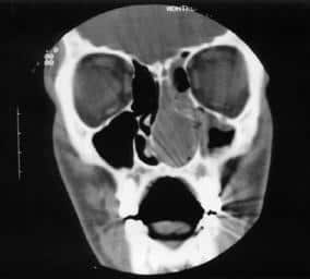
| A. | Antrochoanal polyp |
| B. |
Ethmoidal polyp |
| C. | Nasaopharyngeal Angiofibroma |
| D. |
Inverted papilloma |
Show Answer

|
Correct Answer � C Explanation |
 |
Ans : C.)Nasopharyngeal Angiofibroma
NASOPHARYNGEAL ANGIOFIBROMA
- It is a histologically benign but locally aggressive vascular tumor that grows in the back of the nasal cavity.
- It is the most common benign neoplasm of nasopharynx.
- It is a highly vascular tumor.It consists of fibrous tissue and blood vessels,
- Blood supply of the tumor most commonly arises from the internal maxillary artery.
- The exact cause is unknown. As the tumour is predominantly seen in adolescent males in the second decade of life, it is thought to be testosterone dependent.
- Such patients have a hamartomatous nidus of vascular tissue in the nasopharynx and this is activated to form angiofibroma when male sex hormone appears.
- Most common site is posterior part of nasal cavity close to the margin of sphenopalatine foramen.
- It may expand into the surrounding areas like nasal cavity,sphenoid sinus,pterygopalatine fossa,maxillary sinus,orbit and cranial cavity.
CLINICAL PRESENTATION OF NASOPHARYNGEAL ANGIOFIBROMA
- Sex : Seen almost exclusively in males (testosterone dependent).
- Age : 10-20 years (2nd decade).
- Clinical features: – Most common symptom is profuse and recurrent epistaxis Progressive nasal obstruction
- Other features depending upon the extension of the tumour:
- Denasal speech(Rhinolalai clausa)
- Conductive hearing loss and serous otitis media due to eustachian tube blockage
- Broadening of nasal bridge
- Soft palate may bulge due to a large mass in late stages
- Proptosis
- Frog-face deformity
- Swelling of cheek
- Involvement of cranial nerves II, Ill, IV, VI
INVESTIGATIONS IN A CASE OF NASOPHARYNGEAL ANGIOFIBROMA
- Radiography:it shows a soft tissue shadow in the nasopharynx.
- Lateral view shows forward displacement of the posterior wall of maxilla.
- Angiography and CT Scan with contrast deomonstrate the extent and vascularity of the tumor.
- Biopsy should never be attempted as it may lead to profuse bleeding.
- Instead,Excision biopsy is performed.
TREATMENT OF NASOPHARYNGEAL ANGIOFIBROMA
Surgery
- Surgical excision is the treatment of choice.
- Before surgery at least 2-3 liters of blood should be given.
- Preoperative embolization and estrogen therapy or cryotherapy reduce blood loss in surgery.
- Hypotensive Anesthesia in nasopharyngeal angiofibroma is/are given by Propofol and Phentolamine.
- Approach
- Wilson’s Transpalatine approach—done for tumor confined to nasopharynx.
- Lateral rhinotomy approach—done for large tumors involving, nasal cavity, paranasal sinuses and orbit.
- Other approaches:
- Sardana’s approach – Transpalatine + Sublabial.
- Extended Lateral Rhinotomy
- Intracranial combined with Extracranial.
- Infratemporal fossa approach.
Radiation
- It is sometimes advised to reduce the vascularity of the tumor.
- It may be considered in cases with intracranial extension.
Hormonal Therapy
- It is doubtful if tumour regression occurs with this therapy.
