Question
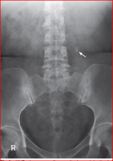
A 52-year-old woman presented with gross haematuria associated with right loin pain for 3 days. On physical examination, she was tender over the right loin region but was afebrile. Blood tests were essentially normal. A KUB was performed as an initial investigation. What can be the most possible diagnosis?
| A. |
Renal Infarct
|
| B. |
Renal Calculus
|
| C. |
Ureteric Calculus
|
| D. |
Renal Carcinoma
|
Show Answer
|
Correct Answer � B
Explanation
|
|
Ans: B.)Renal Calculus
Image shows: Coned KUB shows radioopacities (arrows) over the left renal area which may represent renal stones.
RENAL CALCULUS
-
Renal calculus is a common cause of haematuria, particularly in the presence of loin pain.
-
Renal calculi are composed of crystals and/or aggregated with proteins.
-
The crystal may contain calcium or magnesium oxalate/phosphate (which are radio-opaque and found in the majority of stones) or uric acid/ xanthine (which are not radio-opaque). As a result of this variable composition, the majority (~90%) of renal stones are radio-opaque, hence a KUB is useful as the initial investigation in a patient with suspected urinary calculus.
-
IVU is the key imaging modality for the diagnosis of
-
(1) Suspected urinary tract calculus
-
(2) Radio-lucent calculus which is not visualized on KUB
-
(3) Ureteric calculus may not be demonstrated by ultrasound.
-
Information on IVU which is helpful in the management of patients with renal stone include:
-
If a patient is allergic to an intravenous contrast medium, ultrasound is a good alternative imaging modality for diagnosis.