Question
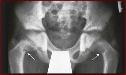
A 45 year old female presented with pain in the pelvic girdle.X ray shows the following features.What can be the most possible diagnosis?
| A. |
Osteogenesis Imperfecta
|
| B. |
Osteopetrosis
|
| C. |
Osteomalacia
|
| D. |
Hypoparathyroidism
|
Show Answer
[ads id=”53026″]
|
Correct Answer � C
Explanation
|
|
Ans:C. Osteomalacia.
Image shows:Looser zones :Symmetric transverse lucent areas in the medial aspect of the femoral necks (arrows): a manifestation of osteomalacia.
OSTEOMALACIA
- Vitamin D deficiency is the most common cause of osteomalacia
- Painful proximal muscle weakness (especially pelvic girdle); bone pain and tenderness.
- Decreased bone density from defective mineralization.
- Decrease in the rate of bone turnover and an increase in the amount of uncalcified osteoid.
Laboratory abnormalities
- Increased alkaline phosphatase
- Decreased 25-hydroxyvitamin D
- Hypocalcemia
- Hypocalciuria
- Hypophosphatemia
- Secondary hyperparathyroidism
Radiological features
- The classical feature: Pseudo fracture or Looser’s zone.
- Triradiate pelvis in females.
- Protrusion-acetabuli: acetabulum protruding into the pelvis.
- More commonly there is diffuse fading of skeleton leading to biconcave (cod fish) vertebra due to disc pressure and indentation of acetabulum producing trefoil or champagne glass pelvis.
Like this:
Like Loading...