Question

| A. |
Brachioradialis |
| B. |
Extensor carpi radialis longus |
| C. |
Flexor carpi radialis |
| D. |
Extensor carpi ulnaris |

|
Correct Answer » A Explanation |
 |
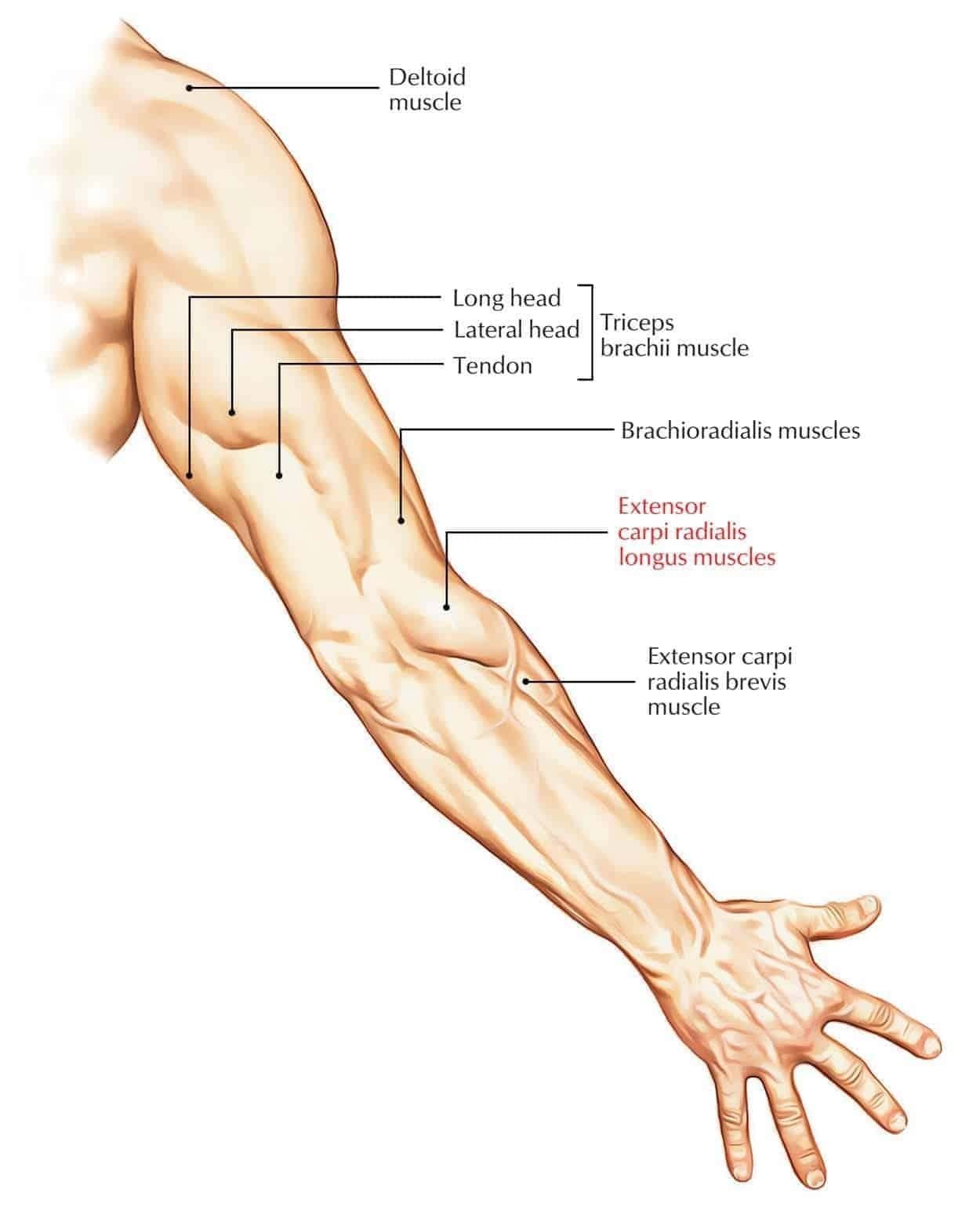
The image highlights a superficial muscle on the lateral side of the forearm.
– The marked muscle emerges from the lateral supracondylar ridge of the humerus and runs along the radial (thumb) side to insert near the styloid process of the radius.
Clinical Reasoning for the Correct Answer:
– The muscle labeled ‘A’ is most prominent when the forearm is in mid-pronation; this matches the position and bulge of the *brachioradialis*.
– *Brachioradialis* originates from the lateral supracondylar ridge of the humerus and inserts at the lateral aspect of the distal radius, just above the styloid process—consistent with the diagram.
Why Option A is Correct (Brachioradialis):
– Located laterally in the forearm.
– Most superficial muscle, forming the lateral border of the cubital fossa.
– Its location, origin, and insertion match the anatomical marking in the image.
Why Option B is Incorrect (Extensor carpi radialis longus):
– Lies just medial (ulnar side) to brachioradialis.
– Does not form the most lateral forearm bulge.
– Its tendon continues to the base of the 2nd metacarpal, not the radius.
Why Option C is Incorrect (Flexor carpi radialis):
– Located in the anterior (flexor) compartment of the forearm.
– Lies more medially and deeper compared to the brachioradialis.
– Not visible on the lateral side in this view.
Why Option D is Incorrect (Extensor carpi ulnaris):
– Located on the ulnar (medial) side of the posterior forearm.
– Forms the medial border of the extensor compartment.
– Far from the lateral aspect where muscle ‘A’ is located.