Question
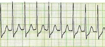
This patient came to the casualty with palpitations. His ECG has been shown below. What is your diagnosis?
| A. |
Ventricular tachycardia
|
| B. |
A-V dissociation
|
| C. |
Supraventricular tachycardia
|
| D. |
Sinus tachycardia
|
Show Answer
|
Correct Answer » C
Explanation
|
|
- ECG shows a rapid regular rhythm.
- R-R interval is approximately 1.5 large boxes.
- Heart rate is around 180–190/min.
- QRS complexes are narrow.
- P waves are not clearly visible.
- This is characteristic of a narrow-complex regular tachycardia, most likely AV nodal re-entrant tachycardia (AVNRT), a common type of SVT.
Supraventricular Tachycardia
- SVT originates above the ventricles.
- AVNRT is the most common type.
- ECG features:
- Regular rhythm
- Narrow QRS complex
- Rate 150–250/min
- P waves absent or retrograde
- Common symptoms:
- Palpitations
- Dizziness
- Chest discomfort
- Dyspnea
- Acute treatment:
- Vagal maneuvers
- IV adenosine
- Beta blockers or calcium channel blockers
- Synchronized cardioversion if unstable
- Features favoring VT instead of SVT:
- Wide QRS complex
- AV dissociation
- Fusion beats
- Structural heart disease
Why Other Options are Incorrect
A. Ventricular tachycardia
- VT usually produces wide QRS complexes (>120 ms).
- AV dissociation, fusion beats, or capture beats may be present.
- Current ECG shows narrow QRS complexes, making VT unlikely.
B. Atrioventricular (AV) dissociation
- AV dissociation is an ECG finding, not a primary rhythm diagnosis.
- It is commonly seen in VT or complete heart block.
- No evidence of independent atrial and ventricular activity is seen here.
D. Sinus tachycardia
- Sinus tachycardia usually has visible upright P waves before every QRS.
- Rate is generally <160/min in adults.
- Sinus tachycardia has gradual onset and termination.
- This ECG shows very rapid regular tachycardia without clear sinus P waves, favoring SVT.