
Question
What is the probable diagnosis
| A. |
Ankylosing spondylitis |
| B. |
Diffuse idiopathic skeletal hyperostosis (DISH) |
| C. |
OA spine |
| D. |
Osteopetrosis |
Show Answer

|
Correct Answer � A Explanation |
 |
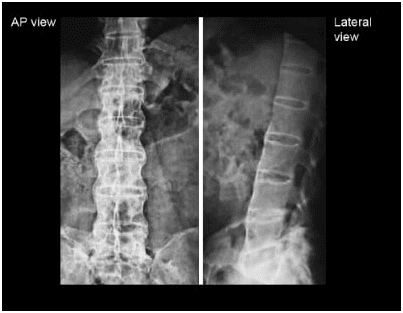
Answer A) Ankylosing spondylitis
Given Image: Involvement of the lumbar spine & the classical Bamboo spine appearance.
Ankylosing spondylitis
Ankylosing spondylitis (less commonly known as Bechterew disease or Marie Strümpell disease) is a seronegative spondyloarthropathy, which results in fusion (ankylosis) of the spine and sacroiliac (SI) joints, although involvement is also seen in large and small joints.
Plain radiograph
Sacroiliac joints
Sacroiliitis is usually the first manifestation and is symmetrical and bilateral
- the sacroiliac joints first widen before they narrow
- subchondral erosions, sclerosis, and proliferation on the iliac side of the SI joints
- at end-stage, the SI joint may be seen as a thin line or not visible
Spine
- early spondylitis is characterized by small erosions at the corners of vertebral bodies with reactive sclerosis: Romanus lesions of the spine (shiny corner sign)
- vertebral body squaring
- noninfectious spondylodiscitis: Andersson lesion
- diffuse syndesmophytes ankylosing can give a “bamboo spine” appearance
syndesmophytes are classically described as paravertebral ossification running parallel to the spine
- linear ossification along the central spine; representing interspinous ligament ossification can give a “dagger spine” appearance on frontal radiographs;
- ossification of spinal ligaments, joints and discs (with fatty marrow within the ossified disc, best seen on MRI)
- apophyseal and costovertebral arthritis and ankylosis
- enthesophyte formation from enthesopathy
- pseudoarthrosis may form at fracture sites
- dural ectasia
