Question
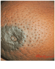
A 35 year old female presented to OPD. Physical examination findings are given in image. Which structure is affected in this condition?
| A. |
Involvement of chest wall
|
| B. |
Involvement of subdermal lymphatics
|
| C. |
Involvement of Cooper’s ligaments
|
| D. |
Involvement of connective tissue
|
Show Answer
|
Correct Answer » B
Explanation
|
|
The image shows:
- Peau d’orange (orange peel appearance) of the breast skin.
- Multiple prominent skin pores with surrounding edema.
- This appearance is highly suggestive of lymphatic obstruction due to carcinoma of the breast, particularly inflammatory breast carcinoma or locally advanced breast cancer.
Clinical Explanation
Peau d’orange occurs when:
- Tumor cells infiltrate and obstruct the subdermal (dermal) lymphatic vessels.
- Lymphatic drainage is impaired, causing cutaneous edema.
- The skin swells, but remains tethered at the hair follicles and sweat gland openings, producing the characteristic orange peel appearance.
Explanation of the Correct Answer
B. Involvement of the subdermal lymphatics
- Obstruction of the subdermal lymphatic channels causes lymphatic edema.
- The edema accentuates the hair follicles and skin pores, producing peau d’orange.
- It is an important clinical sign of breast carcinoma, especially inflammatory carcinoma.
Why Other Options are Incorrect
A. Involvement of the chest wall
- Causes fixation of the breast to the underlying muscles.
- Produces restricted mobility of the breast.
- Does not cause peau d’orange.
C. Involvement of Cooper’s ligaments
- Infiltration of Cooper’s suspensory ligaments causes skin dimpling or puckering.
- It does not produce the diffuse orange peel appearance.
D. Involvement of connective tissue
- This is a nonspecific finding.
- Peau d’orange specifically results from lymphatic obstruction, not generalized connective tissue involvement.