Question
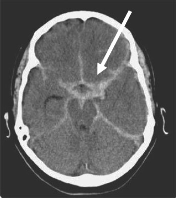
A 40-year-old patient complains of a headache. A CT head was taken, as shown below. What is the most accurate diagnosis?
| A. |
Subarachnoid hemorrhage (SAH) – aneurysm
|
| B. |
Epidural hemorrhage
|
| C. |
Subdural hemorrhage
|
| D. |
Intraparenchymal hemorrhage
|
Show Answer
|
Correct Answer » A
Explanation
|
|
The non-contrast CT shows hyperdensity in basal cisterns, particularly around the circle of Willis—a hallmark of acute subarachnoid hemorrhage (SAH) from an aneurysmal rupture.
– Clinical Presentation:
– Sudden, severe “thunderclap” headache indicates SAH.
Differential CT Features:
– B. Epidural hemorrhage:
– Shape: Biconvex/lens-shaped.
– Location: Between skull and dura, often with skull fracture.
– Distinctive: Not in basal cisterns; associated with trauma.
– C. Subdural hemorrhage:
– Shape: Crescent-shaped along convexity of the brain.
– Location: Between dura and arachnoid.
– Distinctive: Not centered in basal cisterns.
– D. Intraparenchymal hemorrhage:
– Shape: Localized hyperdense area.
– Location: Within brain lobes, basal ganglia, or cerebellum.
– Distinctive: Not in subarachnoid cisterns.
These CT visual cues are critical in differentiating these hemorrhage types.