Question
| A. |
Pineal gland
|
| B. |
Stem of lateral sulcus
|
| C. |
Internal carotid siphon
|
| D. |
Wernicke’s area
|
Show Answer
|
Correct Answer � B
Explanation
|
|
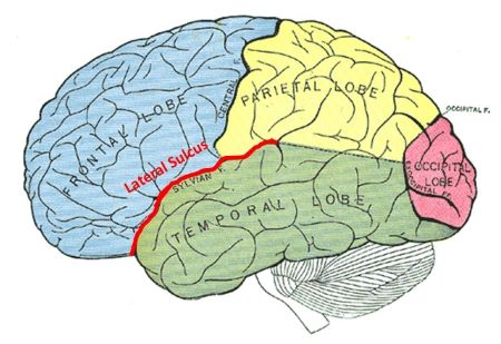
The arrow in the image points to a region associated with the stem of the lateral sulcus (Sylvian fissure), which separates the frontal and parietal lobes from the temporal lobe. This is a key anatomical landmark in the brain.
Key Facts About the Lateral Sulcus:
- Anatomical Location: Separates the temporal lobe from the frontal and parietal lobes.
- Clinical Importance:
- Contains key vascular and neural structures.
- Nearby regions include Broca’s and Wernicke’s areas, important for speech and language processing.
- Branches:
- The posterior ramus extends into the parietal lobe.
- The ascending and anterior rami extend into the frontal lobe.
Image Link : https://medicoapps.org/wp-content/uploads/2024/11/20241119121149.jpg
Explanation of Other Options:
Pineal gland : The pineal gland is located deeper within the brain in the epithalamus, near the roof of the third ventricle. It does not lie in proximity to the lateral sulcus.
Internal carotid siphon: The internal carotid siphon is a part of the internal carotid artery located at the base of the brain. It is not directly visible or related to the lateral sulcus in this image.
Wernicke’s area: Wernicke’s area, associated with language comprehension, is located in the superior temporal gyrus near the posterior part of the lateral sulcus. However, it is not at the specific location indicated by the arrow.