Necrosis
NECROSIS


- Necrosis is defined as localised area of tissue death which later leads to degradation of tissues by hydrolytic enzymes liberated from dead cells.
- Based on morphology 5 types of necrosis-
1. Coagulative necrosis-
- Most common type of necrosis caused by sudden cessation of blood flow.
- It effects- heart, kidney, spleen (except brain).
- Underlying tissue architecture is preserved.
- Coagulative necrosis is characteristics of infarcts in all solid organs (mainly heart).
- Hallmark of coagulative necrosis is conversion of normal cells into their tombstones.
- The cell injury causes denaturation of protein.
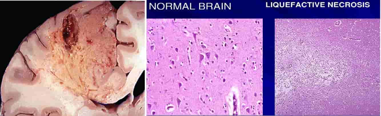
2. Liquefactive necrosis-
- Liquefactive or colliquative necrosis occurs due to lysosomal permeability and enzymes of leukocytes digest the tissue transforming the tissue into liquid viscous mass.
- Tissue architecture is lost.
- Examples are- Infarct brain and abscess cavity.
3. Caseous necrosis-
- Caseous means cheese like appearance.
- It is found in the centre of foci of tuberculous infections & histoplasmosis.
- It has features of both coagulative & liquefactive necrosis.
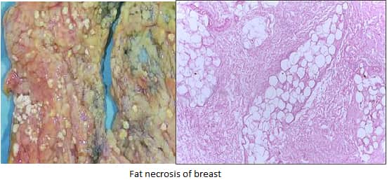
4. Fat necrosis-
- Fat necrosis refers to cell death in fat rich organs of the body.
- Mesentric fat necrosis due to acute pancreatitis as there is liberation of pancreatitic lipases resulting in necrosis of pancreas & peritoneal cavity.
- Traumatic fat necrosis of the breast.
- The released fatty acids combined with calcium gives a chalky white appearance.
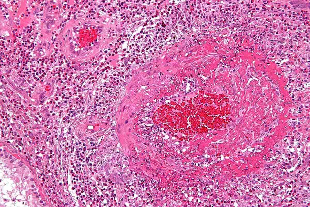
5. Fibrinoid necrosis-
- Fibrinoid necrosis is a special form of necrosis in immune reactions in which antigen and antibodies are deposited on the walls of arteries.
- It is seen in vasculitis & malignant hypertension.
- Seen in sarcoidosis.
Exam Important
Coagulative necrosis-
- It effects- heart, kidney, spleen (except brain).
- Underlying tissue architecture is preserved.
- Coagulative necrosis is characteristics of infarcts in all solid organs (mainly heart).
- Hallmark of coagulative necrosis is conversion of normal cells into their tombstones.
- The cell injury causes denaturation of protein.
- Liquefactive necrosis- Examples are- Infarct brain and abscess cavity.
- Caseous necrosis- It is found in the centre of foci of tuberculous infections & histoplasmosis.
- It has features of both coagulative & liquefactive necrosis.
- Fat necrosis- Mesentric fat necrosis due to acute pancreatitis as there is liberation of pancreatitic lipases resulting in necrosis of pancreas & peritoneal cavity.
- Traumatic fat necrosis of the breast.
- The released fatty acids combined with calcium gives a chalky white appearance.
- Fibrinoid necrosis- Fibrinoid necrosis is a special form of necrosis in immune reactions in which antigen and antibodies are deposited on the walls of arteries.
- It is seen in vasculitis & malignant hypertension.
- Seen in sarcoidosis.
Don’t Forget to Solve all the previous Year Question asked on Necrosis
Click Here to Start Quiz
Click Here to Start Quiz
