
PELVIS MUSCULATURE
| A | Sartorius | |
| B |
Obturator internus |
|
| C |
Obturator externus |
|
| D |
Gluteus minimus |
Muscles that can cause external rotation of the hip include all of the following except:
| A |
Sartorius |
|
| B |
Obturator internus |
|
| C |
Obturator externus |
|
| D |
Gluteus minimus |
Ans:D.)Gluteus Minimus.
Many of the muscles of the gluteal region are external rotators of the hip. These muscles include the gluteus maximus, piriformis, obturator internus, obturator externus, quadratus femoris, superior gemellus, and inferior gemellus. The sartorius is a flexor and external rotator of the hip. The gluteus minimus is an abductor and internal rotator of the hip.
All are sphincters of lower genito urinary tract of female except?
| A |
Pubovaginalis |
|
| B |
External urethral sphincter |
|
| C |
Internal urethral sphincter |
|
| D |
Bulbospongiosus |
Ans. C. Internal urethral sphincter
There is no ‘internal urethral sphincter’ in females. It is present in males at the bladder neck and proximal urethral. Its function is to prevent retrograde ejaculation of semen into the bladder during the time of ejaculation.
In males, the detrusor muscle fibers of the bladder at the region of bladder neck and proximal urethra are organized into internal urethral sphincter which is under involuntary control.
External urethral sphincter is present in both males and females.
Vaginal sphincter is formed by the fibres of pubococcygeus known as pubovaginalis.
There are other muscles also which contribute to compressing the vaginal orifice.
| A |
Pubococcygeus |
|
| B |
Iliococcygeus |
|
| C |
Pubovaginalis |
|
| D |
All of the above |
The pelvic diaphragm forms a broad muscular sling and provides substantial support to the pelvic viscera.
This muscle group is comprised of the levator ani and the coccygeus muscle.
| A |
Colle’s fascia |
|
| B |
Sphincter urethrae |
|
| C |
Perineal membrane |
|
| D |
Deep transverse Perineal muscle |
Urogenital diaphragm is a triangular musculofascial diaphragm situated in the anterior part of perineum, filling in the gap of the pubic arch. It is formed by the sphincter urethrae, deep transverse perineal muscles enclosed between superior and inferior layer of fascia of urogenital diaphragm. Inferior layer of fascia is called perineal membrane.
| A |
Iliococcygeus |
|
| B |
Ischiococcygeus |
|
| C |
Bulbospongiosus |
|
| D |
Levator ani |
Superficial muscles of perineum (lie in superficial perineal pouch) are ischiocavernosus, bulbospongiosus, and superficial transverse perinei.
Deep muscles (lie in deep perineal pouch) are sphincter urethrae, deep transverse perinei, and in females, compression urethrae and sphincter urethrovaginalis.
| A |
Gluteus maximus |
|
| B |
Perineal membrane |
|
| C |
Pelvic diaphram |
|
| D |
Obturator internus |
Ans. is ‘d’ i.e., Obturator internus
Boundries of ischeoanal (ischeorectal) fossa are :-
- Anteriorly :- Posterior border of perineal membrane .
- Posteriorly :- Gluteus maximus muscle, sacrotuberous ligament.
- Laterally :- Ischial tuberosity and obturator internus.
- Medially :- Sphincter ani externus (external anal sphincter) and pelvic diaphragm (levator ani).

| A | Puborectalis is essential to maintain continence. | |
| B |
Internal sphincter is skeletal muscle. |
|
| C | Internal sphincter remains in the state of tonic contraction. | |
| D |
Both A and C. |
Ans;D)Both A and C.
The organ marked by a “red box” in the picture above represents the anal canal.
In the anal canal Puborectalis is essential to maintain continence and Internal sphincter remains in the state of tonic contraction.
Structures crossing the dorsal surface of the given structure marked by a “red arrow” are?

| A |
Internal pudendal vessel. |
|
| B |
Pudendal nerve. |
|
| C |
Obturator nerve. |
|
| D |
Nerve to obturator internus. |
Ans;C).Obturator nerve
The structure marked by a red arrow represents the ischial spine.
ISCHIAL SPINE-
A thin pointed triangular eminence that projects from the dorsal border of the ischium and gives attachment to the gemellus superior on its external surface and to the coccygeus, levator ani, and pelvic fascia on its internal surface
The structure crossing the dorsal surface of the ischial spine is the Obturator nerve.
Psoas major, iliacus & pectineus muscles, femoral vessels and nerve, the femoral branch of the genitofemoral nerve, lateral cutaneous nerve of thigh and lymphatics pass below the inguinal ligament.
‘PIN’ structures i.e. Pudendal nerve, Internal Pudendal vessels, Nerve to obturator internus come out of greater sciatic foramen, cross the dorsal surface of ischial spine & enter into the lesser sciatic foramen.
From the posterior border of the body of the Ischium there extends backward a thin and pointed triangular eminence, the ischial spine, more or less elongated in different subjects.
Clinical significance
It can serve as a landmark in pudendal anesthesia
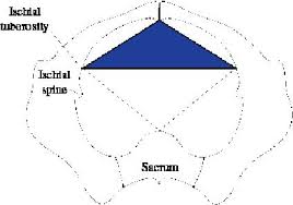
All of the following structures forms the boundary of the triangle as shown in the picture below, EXCEPT ?
| A |
Pubic rami. |
|
| B |
Ischial tuberosity. |
|
| C |
Ischio cavernosus muscle. |
|
| D |
Superficial transverse perineal muscle. |
The triangle shown in the picture above represents Urogenital triangle.
The perineum is divided by an arbitrary line joining the ischial tuberosities into urogenital triangle and anal triangle. The urogenital triangle is bounded by pubic rami superiorly, the ischial tuberosities laterally, and the superficial transverse perineal muscle posteriorly.
All of the following are true about sphincter urethrae except?
| A | Voluntary | |
| B |
Supplied by pudendal nerve |
|
| C |
Arises from ischiopubic ramus |
|
| D |
Located at bladder neck |
Ans. d. Located at bladder neck
External urethral sphincter
Voluntary
Made of striated muscle fibres
It is roughly pear shaped, being thickest near the middle of urethra and thicker in front than at sides or back.
Supplied by perineal branch of pudendal nerve (S2-S4)
Origin: From ischiopubic rami on each side
Internal urethral sphincter
Involuntary
Made of smooth muscle
Continuation of detrusor muscle
It is an extension of circular muscle at the bladder neck
Supplied by sympathetic nerves (T10 -L1)
Females lack internal urethral sphincter
Support of prostate is ‑
| A |
Pubococcygeus |
|
| B |
Ischiococcygeus |
|
| C |
Ilioccygeus |
|
| D |
None of the above |
Ans. is ‘a’ i.e., Pubococcygeus
- Levator ani muscle is divisible into following parts :1) Puboccygeus part 2)Iliococcygeus
Pubococcygeus runs backward from the body of pubis toward the coccyx. Some of the fibres get inserted into prostate, urethra, vagina, and anorectal junction and named accordingly : Puboprostaticus , pubourethralis, pubovaginalis, puborectalis respectively.
Superficial perineal space contains ‑
| A |
Sphincter urethrae muscle |
|
| B |
Ischiocavernosus muscle |
|
| C |
Deep transverse perinei muscle |
|
| D |
Bulbourethral gland |
Ans. is ‘b’ i.e., Ischiocavernosus muscle
