PLACENTA
The placenta is developed from two sources:
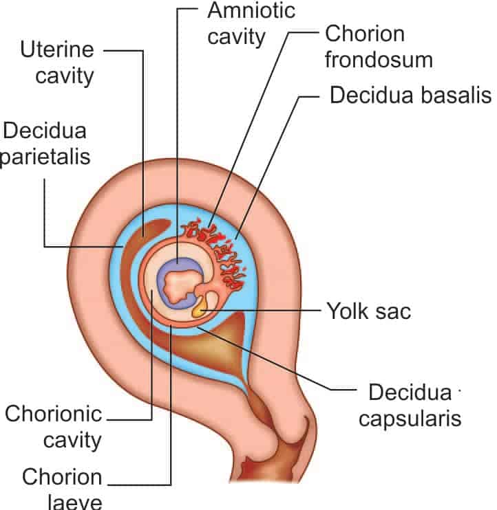
- Fetal(Develops from the chorion frondosum)
- Maternal (consists of decidua basalis)
Interstitial implantation(11th DAY)
↓(Blastocyst surrounded on all sides by lacunar spaces)
Trabeculae ↓
↓Multilocular receptacle lined by syncytium and filled with maternal blood→ xIntervillous space.
Stem villi(13th DAY)
↓(connect chorionic plate with basal plate)
Primary, secondary and tertiary villi
↓(In mesenchymal core of villi)
Arterio-capillary-venous system(21st DAY)
↓Connects intraembryonic vascular system through the body stalk
At 6th week and both the villi and the lacunar spaces in the abembryonic area get obliterated compensated by:
- Decidua basalis(exuberant growth and proliferation) +
- Chorion frondosum(Exuberant division and subdivision of the chorionic villi in embryonic pole) ↓
Discrete placenta (From 6th week to 12th week)


ANATOMY:
- Circular disk
- Diameter :15–20 cm
- Weighs:500 gm(weight of fetus to weight of placenta is 6 : 1)
- Two surfaces, fetal and maternal, and a peripheral margin.
Fetal surface:
- Covered by amnion
- Umbilicalcord attached at or near its center
- Branches of the umbilical vessels are visible beneath the amnion
- 4/5 of total placenta.
Maternal surface:
- Remnant of the decidua basalis
- Lobes or cotyledons(15–20 convex polygonal areas limited by fissures.)
- Each fissure is occupied by the decidual septum .
- Grayish spots: Calcium deposits
- 1/5 of total placenta
- Decidua basalis and blood in the intervillous space are of maternal origin
Peripheral margin:
- Limited by the fused basal and chorionic plates
- Continuous with the chorion laeve and amnion
Attachment:
- Upper part of the body of the uterus encroaching to fundus adjacent to the anterior or posterior wall
Line of separation:
- Decidua spongiosum
The placenta consists of two plates:
Chorionic plate:
- Lined by the amniotic membrane.
- The umbilical cord is attached to this plate
From within outward structures:
- Primitive mesenchymal tissue containing branches of umbilical vessels
- cytotrophoblast
- syncytiotrophoblast.
Basal plate
- Lies to the maternal aspect
- Between the two plates lies the intervillous space containing the stem villi
- The space being filled with maternal blood
Structures from outside inwards:
- Decidua basalis
- Nitabuch’s layer of fibrinoid degeneration of the outer syncytiotrophoblast
- Cytotrophoblastic shell
- Syncytiotrophoblast
AMNIOTIC MEMBRANE:
- Cubical epithelium
- Attached to chorionic plate
INTERVILLOUS SPACE:
Boundary:
- Inner:chorionic plate
- Outer:basal plate
- Lining:syncytiotrophoblast
- Filling:maternal blood
- Numerous branching villi from the stem villi project into the space
STEM VILLI:
- Functional unit of the placenta is called a fetal cotyledon or placentome, is derived from a major primary stem villus
- Functional subunit:lobule,derived from a tertiary stem villi
- About 60 stem villi persist in human placenta.
- Each cotyledon (total 15–29) contains 3–4 major stem villi
STRUCTURE OF A TERMINAL VILLUS:
- In the early placenta:Outer syncytiotrophoblast→ cytotrophoblast → basement membrane→ Central stroma containing fetal capillaries, primitive mesenchymal cells, connective tissue & phagocytic (Hofbauer) cells
In placenta at term:
- Site for transfer: Syncytiotrophoblast thin overlying fetal capillaries
- Site for synthesis: Thick Syncytiotrophoblast containing extensive endoplasmic reticulum
- Basement membrane becomes thicker
- Stroma:Dilated vessels,mesenchymal cells, connective tissue & phagocytic (Hofbauer) cell

- Transfer of nutrients and waste products between the mother and fetus
Endocrine function:
- Insulin, steroids from the adrenals, thyroid, chorionic gonadotrophin or placental lactogen cross the placenta
- Progesterone production require fetal steroidogenic tissue
- Parathormone & calcitonin not crosses placenta
Barrier function:>500 daltons are held up
- HBV viruses is least likely to cross placenta
- Trophoblast, Fetal capillary endothelium & Mesoderm are constituent of placental barrier
Immunological function:
- Placental hormones:Immunosuppressive effect(proteins (SP1), early pregnancy factor (EPF), PAPP-A, Chorionic thyrotropin & chorionic corticotropin and chorionic gonadotropin)
- Human chorionic gonadotropin is produced by the human placenta having LH-like activity
- Extravillous trophoblast:express HLA Class I molecules
- Human chorionic somatotropin (HCS):stimulation of ductal growth in the mammary gland during pregnancy
- Shift of maternal response from cell-mediated (T helper 1) to humoral (T helper 2) immunity
Exam Important
- Human chorionic gonadotropin is produced by the human placenta having LH-like activity
- The placental hormone that participates in stimulation of ductal growth in the mammary gland during pregnancy is Human chorionic somatotropin (HCS)
- HBV viruses is least likely to cross placenta
- Placenta develops from Placenta frondosum & Decidua basalis
- Trophoblast, Fetal capillary endothelium & Mesoderm are constituent of placental barrier
- At term, ratio of weight of fetus to weight of placenta is 6 : 1
- Syncytiotrophoblast is the inner most part of placenta
- In the placenta, maternal blood comes in direct contact with syncytiotrophoblast
- Chorionic gonadotropin, Chorionic thyrotropin & chorionic corticotropin are placental hormones
- Progesterone production require fetal steroidogenic tissue
- Weight of placenta at term is 500 grams
Click Here to Start Quiz
