Plasmodium: Diagnosis
Laboratory Diagnosis
- Microscopy – detecting & identifying malarial parasites in peripheral blood films.
- Concentrating parasites in venous blood by centrifugation when they cannot be found in blood films
- Using a rapid malaria Ag or enzyme detection test
- Other tests – Hb, PCV, Blood glucose, total WBC & platelet count.
Examination of blood Film
- Collection of blood
- Best prepared directly from capillary blood
- In EDTA bulb (used within 30 mins)
- Time of collection
- As soon as possible if malaria is suspected
- Before administering antimalarials
- During pyrexia lphase
Types of blood Film
- Two types:
1.Thick films :
- 30 to 40 times more sensitive than thin films
- More suitable for detection of malarial parasite when they are few in number
- Blood is not fixed, RBCs are lysed during staining (only parasitic forms will be seen)
2. Thin films :
- To confirm the Plasmodium species
- Assists in the identification of mixed infections
- Blood is fixed, parasites are seen within the RBCs
- Also helps in assessing the response to treatment.
Fixation and Staining
- Fixation – thin films are fixed with absolute alcohol for 1 to 2 mins.
- Staining – films are stained with Romanowsky stain: Giemsa, field’s, Wright’s
- Giemsa – 10% solution for 10 mins
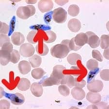
Identifying Blood Film
Look for the different morphological forms of the parasite in blood smear:
1.Trophozoites / ring forms
2.Schizont(Not seen in Plasmodium falciparum)
3.Gametocytes
Trophozoites / ring forms
| Character | P. vivax | P. falciparum |
| Size | 2.5µ (1/3rd of RBC) | 1.25 to 1.5 µ |
| Cytoplasm | Thick opposite to nucleus | Uniform thickness |
| Nucleus | One/ ring | Can have >1 |
| Number of rings | One ring/ RBC | >1/ RBC |
| Location in RBCs | Always inside RBCs | Inside as well as on the surface (accole’ forms) |
| Type of RBC infected | Preferentially young RBCs & reticulocytes | All types |
Schizont
| Character | P. vivax | P. falciparum |
| Size of RBC | Increases to twice its size | Does not change |
| No of merozoites | 16 | 8 to 32 |
| Arrangement of merozoites | Symmetric in form of rosette | Asymmetrical |
| Presence in peripheral blood | Present | Absent |
Gametocytes
| Character | P. vivax | P. falciparum |
|
Shape – Male
Female |
Spherical
Spherical |
Cresentic
Sausage shaped |
|
Nucleus – M
F |
Central, diffuse
Peripheral,small |
Central, diffuse
Central,compact |
| Infected RBC | Enlarged | Deformed, with its membrane, stretched. |
Important Points
Schüffner’s dots
-
Exclusively found in Plasmodium ovale and Plasmodium vivax
- Morphologic alterations in infected host erythrocytes that are visible as multiple brick-red dots
Maurer’s dot:
- Fine granular precipitates or irregular cytoplasmic particles present in red blood cells infected with the trophozoites of Plasmodium falciparum
Counting the % age of parasitised RBCs
- On thin blood films
- When falciparum malaria parasitemia is high
Method of counting:
- Select an area where no of RBCs is roughly 250.
- Count the no of parasitised RBCs in 4 such fields i.e. approximately 1000 RBCs.
- Divide by 10 to obtain the percentage.
*WHO – if it is >5%, then the parasitemia is heavy & prognosis is poor.
Rapid Diagnostic Test
- Developed to diagnose falciparum malaria rapidly & without a microscope.
- Can also detect vivax malaria
- Immunochromatographic test
- Three tests are available commercially
- Detects HRP2 Ag (Histidine-rich protein) or specific pLDH (parasite lactate dehydrogenase) or Pan malarial plasmodium aldolase
- Both HRP2 & pLDH are produced by the parasites during their growth & differentiation in RBCs.
Exam Important
Laboratory Diagnosis
- Microscopy – detecting & identifying malarial parasites in peripheral blood films.
- Concentrating parasites in venous blood by centrifugation when they cannot be found in blood films
- Using a rapid malaria Ag or enzyme detection test
- Other tests – Hb, PCV, Blood glucose, total WBC & platelet count.
Thin films :
- To confirm the Plasmodium species
Identifying Blood Film
Look for the different morphological forms of parasite in blood smear:
- Trophozoites / ring forms
- Schizont(Not seen in Plasmodium falciparum)
- Gametocytes
Schüffner’s dots
-
Exclusively found in Plasmodium ovale and Plasmodium vivax
- Morphologic alterations in infected host erythrocytes that are visible as multiple brick-red dots
Maurer’s dot:
- Fine granular precipitates or irregular cytoplasmic particles present in red blood cells infected with the trophozoites of Plasmodium falciparum
Immunochromatographic test
- Three tests are available commercially
- Detects HRP2 Ag (Histidine-rich protein) or specific pLDH (parasite lactate dehydrogenase) or Pan malarial plasmodium aldolase
- Both HRP2 & pLDH are produced by the parasites during their growth & differentiation in RBCs.
Don’t Forget to Solve all the previous Year Question asked on Plasmodium: Diagnosis
Click Here to Start Quiz
Click Here to Start Quiz
